Review
. 2012 Jan 4:709:21-31.
doi: 10.1016/j.aca.2011.10.022. Epub 2011 Oct 19.

Protein separation by capillary gel electrophoresis: a review

Affiliations
Review

Protein separation by capillary gel electrophoresis: a review

Zaifang Zhu et al. Anal Chim Acta. .

Abstract

Capillary gel electrophoresis (CGE) has been used for protein separation for more than two decades. Due to the technology advancement, current CGE methods are becoming more and more robust and reliable for protein analysis, and some of the methods have been routinely used for the analysis of protein-based pharmaceuticals and quality controls. In light of this progress, we survey 147 papers related to CGE separations of proteins and present an overview of this technology. We first introduce briefly the early development of CGE. We then review the methodology, in which we specifically describe the matrices, coatings, and detection strategies used in CGE. CGE using microfabricated channels and incorporation of CGE with two-dimensional protein separations are also discussed in this section. We finally present a few representative applications of CGE for separating proteins in real-world samples.


Figures

Fig. 1
Fig. 1. Immunoblot chip
(a) Schematic design of the immunoblot chip for analysis of native proteins. The sample (2), sample waste (3), buffer (1, 4, 5, 6) and buffer waste (7, 8) reservoirs are indicated in sketch (not to scale). The middle region of the device (indicated as Chamber) has a length of 1.5 mm, a width of 1 mm and a depth of 20 µm. (b) Three separate zones inside the Chamber to facilitate protein immunoblotting: a large-pore-size protein loading gel on the top, a smaller-pore-size protein separation gel on the bottom-left and an antibody-functionalized blotting gel on the bottom-right. Colored dyes were used to visualize the different gel regions. Reprinted from ref. [85] with permission.
Fig. 2
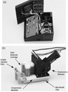
Fig. 2. µChemLab™ instrument
(a) The µChemLab instrument with the top off showing the separation platform, the control panel, the back of the LCD display, and the battery pack. The instrument is approximately 7″×8″×4.5″ and weighs 6 lbs. (b) The separation platform houses the microfluidic chip in a compression manifold that connects the chip to eight fluid reservoirs, two sample injection ports, and a LIF detection module. The overall size of the platform is approximately 5″×3″×4″. Reprinted from ref. [48] with permission.
Fig. 3
Fig. 3. Schematic of a plastic microchip for 2-D protein separation
Reprinted from ref. [90] with permission.
Fig. 4
Fig. 4. CIEF Integrated with CGE in a linear format
(A) Overall Arrangement of experimental setup. (B) Dialysis hollow fiber interface: (1) methacrylate plate, (2) capillaries, (3) Teflon tubes, (4) hollow fiber and (5) buffer reservoir. Reprinted from ref. [98] with permission.
Fig. 5
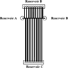
Fig. 5. Microchip for 2D protein separation
(A) Geometrical layout of the microchip used for SDS-µCGE-MEKC. (B) Fluorescence image of the sieving matrix/MEKC interface at the intersection of the SDS-µCGE and MEKC dimensions. Reprinted from ref. [92] with permission.
Fig. 6
Fig. 6. Schematic demonstration of pseudo-SDS dye-protein-SDS complex
Reprinted from ref. [114] with permission.
Fig. 7
Fig. 7. Schematic diagram of experimental apparatus for SDS-CGE and protein collection
(a) SDS-CGE setup with membrane collectror; (b) split view of membrane collector. Reprinted from ref. [117] with permission.

Similar articles

Cited by

References

    1. Shapiro AL, Vinuela E, Maizel JV. Biochem. Biophys. Res. Commun. 1967;28:815–820. - PubMed
    1. Weber K, Osborn M. J. Biol. Chem. 1969;244:4406–4412. - PubMed
    1. Guttman A, Nolan J. Anal. Biochem. 1994;221:285–289. - PubMed
    1. Shieh PCH, Hoang D, Guttman A, Cooke N, J Chromatogr A. 1994;676:219–226.
    1. Guttman A. TrAC Trends Anal. Chem. 1996;15:194–198.

Publication types

MeSH terms

LinkOut - more resources