. 2011 Nov 28;5(1):74.
doi: 10.1186/1752-153X-5-74.

Chemical characterization of some substituted hydroxyapatites

Affiliations

Chemical characterization of some substituted hydroxyapatites

Doreya Mohamed Ibrahim et al. Chem Cent J. .

Abstract

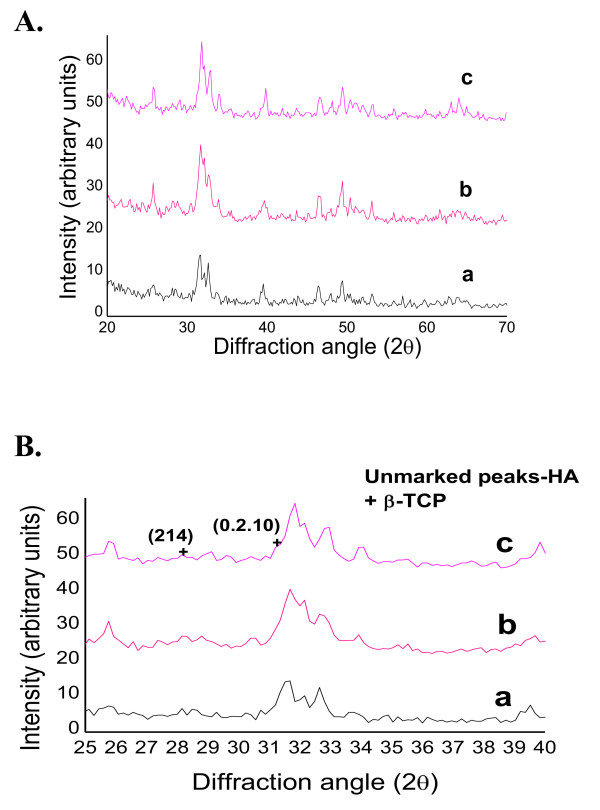
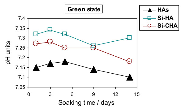
Synthetic multi-substituted hydroxyapatite nano powders containing silicon and or carbonate prepared by a wet chemical method. The process parameters are set up to allow the simultaneous substitution of carbonate and silicon ions in the place of phosphorus. The chemical and structural characterizations of the prepared powders are determined with the aid of; XRF, ICP, XRD and FTIR. The results show that, the ion substitution in the crystal lattice of HA caused a change in the unit cell dimensions and affected the degree of crystallization of the produced powders. The apatite formation abilityy of the prepared discs from the synthesized powders is determined by immersing in SBF solution for different periods. The degree of ion release was determined in the obtained solutions. The examined surface of the immersed discs under SEM and analyzed by CDS showed a more dense HA layer than those of un-substituted ones. The HA with the substituted silicon and carbonate ions, showed the highest solubility with greater rate of ion release, compared with carbonate-free powder. All prepared powders took sodium ion from the SBF solution during immersion, which was not recorded before.


Figures

Figure 1
Figure 1
Flow chart of the method for HA preparation.
Figure 2
Figure 2
XRD spectra of the investigated multi-substituted HA green: (a, HAs); (b, Si-HA); (c, Si-CHA).
Figure 3
Figure 3
XRD spectra of the investigated multi-substituted HA fired at 900°C: A. (a, HAs); (b, Si-HA); (c, Si-CHA). B. (a, HAs); (b, Si-HA); (c, Si-CHA).
Figure 4
Figure 4
IR spectra of multi-substituted HA as-prepared powders: (a, HAs); (b, Si-HA); (c, Si-CHA).
Figure 5
Figure 5
IR spectra of multi-substituted HA powders fired at 900°C: (a, HAs); (b, Si-HA); (c, Si-CHA).
Figure 6
Figure 6
Changes in SBF pH with time of immersion for the green discs of different hydroxyapatites. pH of control SBF at ~ 7.4.
Figure 7
Figure 7
Changes in SBF pH with time of immersion for the fired discs of different hydroxyapatites. pH of control SBF at ~ 7.4.
Figure 8
Figure 8
Calcium ion concentration in simulated body fluid of HAs and the two substituted-HA green samples. Calcium ion concentration of SBF: 10 mg/dl.
Figure 9
Figure 9
Calcium ion concentration in simulated body fluid of HAs and the two substituted-HA heat-treated samples. Calcium ion concentration of SBF: 10 mg/dl.
Figure 10
Figure 10
Change in phosphorus ion concentration of SBF solution with immersion period of green discs for different hydroxyapatites. Phosphorus ion concentration of SBF: 4.33 mg/dl.
Figure 11
Figure 11
Change in phosphorus ion concentration of SBF solution with immersion period of fired discs for different hydroxyapatites. Phosphorus ion concentration of SBF: 4.33 mg/dl.
Figure 12
Figure 12
Change in sodium ion concentration in SBF with immersion period for green discs of different hydroxyapatites. Concentration of Na ion in SBF: 326.458 mg/dl.
Figure 13
Figure 13
Change in sodium ion concentration in SBF with immersion period for fired discs of different hydroxyapatites. Concentration of Na ion in SBF: 326.458 mg/dl.
Figure 14
Figure 14
Change in silicon ion concentration of green disc with immersion period of hydroxyapatites containing silicon.
Figure 15
Figure 15
Change in silicon ion concentration of fired disc with immersion period of hydroxyapatites containing silicon.
Figure 16
Figure 16
SEM of green discs before and after immersion in SBF solution for 14 days. a) HAs, b) Si-HA, c) Si-CHA.
Figure 17
Figure 17
SEM of fired discs before and after immersion in SBF solution for 29 days. a) HAs, b) Si-HA, c) Si-CHA.
Figure 18
Figure 18
SEM of fired hydroxyapatite disc samples after immersion in SBF for 29 days. a) Si-HA, b) Si-CHA, with higher magnification, × 10000.

Similar articles

Cited by

References

    1. Suchanek W, Yoshimura M. Processing and properties of hydroxyapatite-based biomaterials for use as hard tissue replacement implants. J Mate Res. 1998;13:94–117. doi: 10.1557/JMR.1998.0015. - DOI
    1. Bigi A, Cojazzi G, Panzavolta S, Ripamonti A, Roveri N, Romanello M, Noris Suarez K, Moro L. Chemical and structural characterization of the mineral phase from cortical and trabecular bone. J Inorg Biochem. 1997;68:45–51. doi: 10.1016/S0162-0134(97)00007-X. - DOI - PubMed
    1. Driessens FCM. The mineral in bone, dentin and tooth enamel. Bull Soc Chim Belg. 1997;89:663–689.
    1. Smith DK. In: Hydroxyapatite and Related Materials. Brown PW, Constantz B, editor. Boca Raton, FL: CRC Press; 1994. Calcium Phosphate Apatites in Nature; pp. 29–45.
    1. Carlisle EM. Silicon: a possible factor in bone calcification. Science. 1969;167:279–80. - PubMed

LinkOut - more resources