. 2012 Mar 1;363(1):1-14.
doi: 10.1016/j.ydbio.2011.11.002. Epub 2011 Nov 12.

Smooth muscle α actin is specifically required for the maintenance of lactation

Affiliations

Smooth muscle α actin is specifically required for the maintenance of lactation

Nate Weymouth et al. Dev Biol. .

Abstract

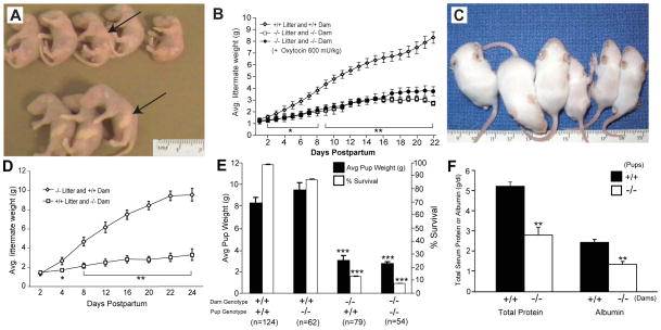
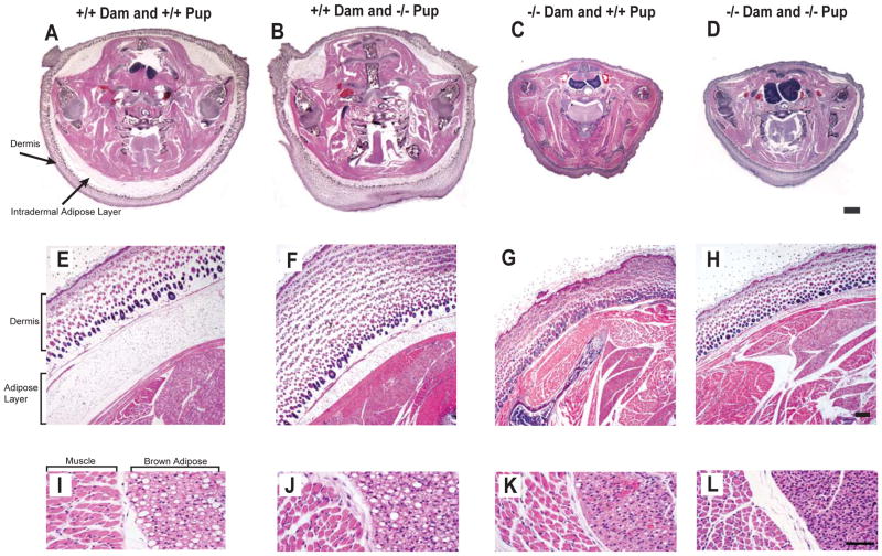
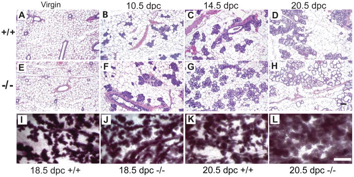
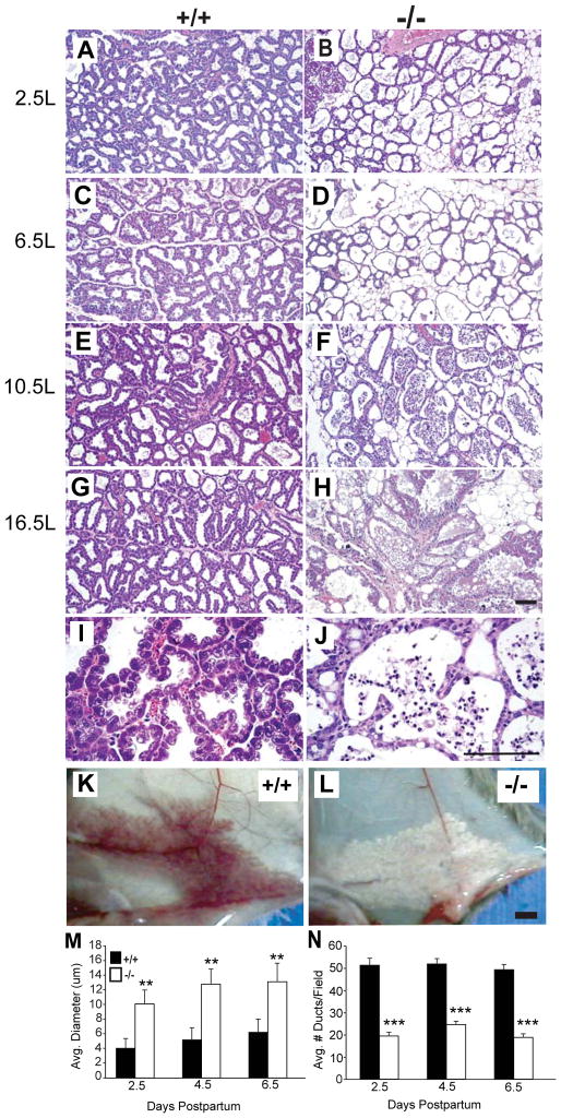
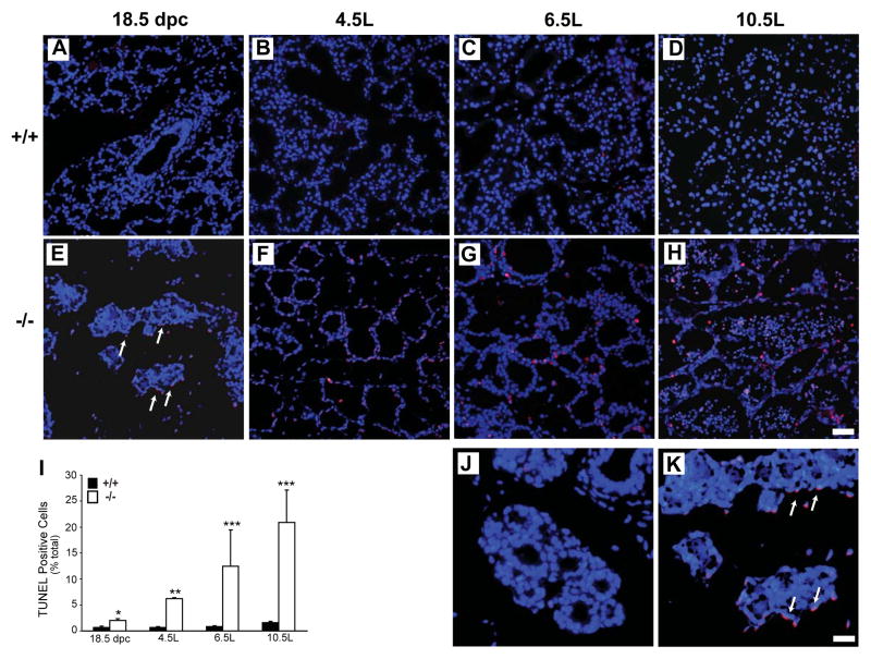
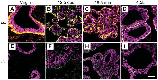
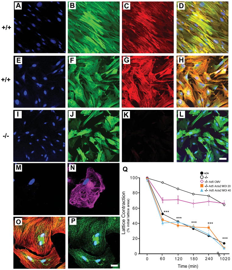
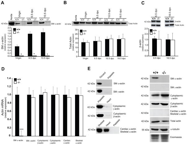
Smooth muscle α-actin (Acta2) is one of six highly conserved mammalian actin isoforms that appear to exhibit functional redundancy. Nonetheless, we have postulated a specific functional role for the smooth muscle specific isoform. Here, we show that Acta2 deficient mice have a remarkable mammary phenotype such that dams lacking Acta2 are unable to nurse their offspring effectively. The phenotype was rescued in cross fostering experiments with wild type mice, excluding a developmental defect in Acta2 null pups. The mechanism for the underlying phenotype is due to myoepithelial dysfunction postpartum resulting in precocious involution. Further, we demonstrate a specific defect in myoepithelial cell contractility in Acta2 null mammary glands, despite normal expression of cytoplasmic actins. We conclude that Acta2 specifically mediates myoepithelial cell contraction during lactation and that this actin isoform therefore exhibits functional specificity.


Conflict of interest statement

Competing interests statement

The authors declare no competing financial interests.

Figures

Fig. 1
Fig. 1. Postnatal growth characteristics of Acta2 +/+ and −/− pups
In (A), is shown a representative example of Acta2 −/− pups nursed by Acta2 −/− dams (upper row) and Acta2 +/+ pups nursed by Acta2 +/+ dams at 4 days postpartum (lower row). Note the absence of milk in the stomach of Acta2 −/− pups and the presence of milk in the Acta2 +/+ pups nursed by Acta2 +/+ dams (arrows point to stomachs). In (B), is shown a graphic summary of postpartum growth of Acta2 +/+ and −/− mice. In addition, Acta2 −/− pups nursed by Acta2 −/− dams were given oxytocin daily in an attempt to stimulate milk letdown (*p<0.05, 2–8 days; **p<0.01: (Acta2 +/+ pups (n=124) vs. Acta2 −/− pups (n=54) or Acta2 −/− pups nursed by Acta2 −/− dams given oxytocin (n=55), 9–22 days). In (C) and (D), Acta2 +/+ and −/− pups were cross fostered to dams of the opposite genotype on day one postpartum. In (C), is shown a representative example; the three pups on the left are Acta2 −/− pups cross fostered with a Acta2 +/+ dam and the three pups on the right are Acta2 +/+ pups cross fostered with an Acta2 −/− dam (day 13 postpartum). In (D), the weights of pups from multiple cross fostering experiments are depicted graphically (*p<0.05, day 4; **p<0.01: Acta2 −/− pups (n=62) vs. Acta2 +/+ pups (n=72), 8–24 days). In (E), survival and postnatal growth at day 22 postpartum of all pups fostered is depicted graphically (***p<0.001: pups nursed by Acta2 −/− vs. Acta2 +/+ dams). In (F), levels of circulating proteins and serum albumin in pups nursed by Acta2 +/+ or −/− dams (n=10; ** p<0.01: pups nursed by Acta2 −/− dams vs. Acta2 +/+ dams).
Fig. 2
Fig. 2. Severe malnourishment in pups nursed by Acta2 −/− dams
Each column contains three pictures from a specific cross fostering pair as indicated on top of each column. In (A–D), pups of the specified genotype and cross fostering dam were sacrificed at day 4 postpartum; midthorax sections are shown (scale bar=1 mm). In (E–H), are representative views of the outer dermis (scale bar= 200 μm) and in (I–L) are representative images of brown adipose regions as indicated (scale bar=50 μm). Note the smaller midthorax diameter (C and D), the reduced layer of adipose fat between the outer dermis and the musculature (G and H) as well as the absence of lipid droplets (K and L) within regions of brown adipose tissue of pups nursed by Acta2 −/− dams.
Fig. 3
Fig. 3. Gestational mammary glands in Acta2 −/− dams appear structurally normal
In (AH), are shown representative examples of mammary glands from Acta2 +/+ (A–D) or −/− (E–H) dams at different gestational time points (H&E, scale bar=100 μm). In (I–L), are shown whole mounts of representative mammary glands at days 18.5 and 20.5 of gestation (scale bar=1 mm). Abbreviation: dpc = days post-coitus.
Fig. 4
Fig. 4. Mammary glands of Acta2 −/− mice exhibit milk stasis, alveolar distension, and precocious involution during lactation
In (A–H), mammary glands were harvested at the indicated times during lactation (H&E, scale bar= 50 μm). Indicated days precede either days post-coitus (dpc) or lactation (L). (I and J) are higher magnification views of the same mammary tissue shown in (E) and (F), respectively (H&E, scale bar= 200 μm). In (K and L), are images of representative mammary glands 12 hours after parturition: an Acta2 +/+ dam (K) and Acta2 −/− dam (L) (scale bar=1 mm). In (M), the alveolar diameter from multiple sections was quantified as in the Methods and the data depicted graphically at the indicated times after parturition (n=5 fields and 6 mice per time point; **p<0.01: Acta2 +/+ dams vs. Acta2 −/− dams for all time points). In (N), ducts were counted as described in the Methods and the average number of ducts per field is depicted (n=5 fields and 6 mice per time point; ***p<0.001 Acta2 +/+ dams vs. Acta2 −/− dams at all time points).
Fig. 5
Fig. 5. Enhanced apoptosis in Acta2 −/− mammary glands
In (A–H), Acta2 +/+ and −/− mammary glands were harvested at the indicated time points. Tissues were subjected to TUNEL as in the Methods. Merged images of DAPI stained nuclei and TUNEL positive cells are shown (DAPI/blue, TUNEL positive cells/pink, scale bar=50 μm). Arrows point to apoptotic cells which lie outside of ducts, surrounding the acini, during late gestation. Indicated days precede either days post-coitus (dpc) or lactation (L). In (I), the ratio of TUNEL positive per DAPI positive cells, per field, are depicted (*p<0.05 Acta2 +/+ dams vs. Acta2 −/− dams at 18 dpc; **p<0.01 Acta2 +/+ dams vs. Acta2 −/− dams at 4L; ***p<0.001 Acta2 +/+ dams vs. Acta2 −/− dams at 6L and 10L; n=6 mice per group and 10 random fields per mouse) In (J and K), are shown higher magnification images of A and E respectively (J- Acta2 +/+, K- Acta2 −/−, scale bar=100 μm)
Fig. 6
Fig. 6. Mammary myoepithelial cell numbers in Acta2 −/− dams are reduced during gestation and lactation
Mammary gland tissue sections from Acta2 +/+ and −/− dams were immunolabeled with anti-cytokeratin 14 (Alexa Fluor 488/green), anti-Acta2 (Cy3/red), and anti-cytokeratin 8 (Cy5/pink) antibodies as in the Methods. In (A–D), are shown representative merged images of stained Acta2 +/+ mammary glands at the indicated time points (scale bar=20 μm). In (E–I), are shown representative merged images of stained Acta2 −/− mammary glands at the indicated time points (scale bar=20 μm). Figures S2 and S3 contain a complete representation of individual staining:(cytokeratin 14 (green), Acta2 (red), and cytokeratin 8 (pink)). Indicated times are symbolized: the days postpartum proceed the letter “L” which stands for lactation. Abbreviation: Dpc = days post-coitus.
Fig. 7
Fig. 7. Phenotypic characterization and rescue of impaired myoepithelial contractility in Acta2 −/− cells
Myoepithelial cells from Acta2 +/+ and −/− dams were isolated as in the Methods. In (A–L), after two days in culture, myoepithelial cells were subjected to immunolabeling to detect the following: sm22α (B, Alexa Fluor 488/green), cytokeratin 14 (F and J; Alexa Fluor 488/green), and smooth muscle α-actin (C, G, and K; Cy3/red). In (D, H, and L) are shown merged images of sm22α or cytokeratin 14 and smooth muscle α-actin immunolabeling (scale bar=50 μm). Control PtK2 epithelial cells were immunolabeled with Acta2 (M, Cy3/red) or cytokeratin 8 (N, Cy5/pink). Control smooth muscle A7r5 cells in (O) were dual immunolabeled with smooth muscle α-actin (Cy3/red) and cytokeratin 14 (Alexa Fluor 488/green). In (P), A7r5 cells were dual immunolabeled with cytokeratin 8 (Cy5/pink) and cytokeratin 14 (Alexa Fluor 488/green) (scale bar=20 μm). Merged images for (O and P) are shown. In (A, E, I, D, H, L, and M–P), BOPRO1/blue was used to label nuclei. In (Q), isolated myoepithelial cells were subjected to collagen lattice contraction assays as described in Methods. In some experiments, isolated Acta2 −/− myoepithelial cells were infected with an adenovirus expressing smooth muscle α-actin (Ad5 Acta2) at the indicted MOIs as in methods (2nd : n=4 for each time point, ***p<0.001 for Ad5 Acta2 treated cells (MOI 20 or 40) vs. Acta2 −/− cells; 1st: n=6 for each time point, ***p<0.001 for Acta2 +/+ cells vs. Acta2 −/− cells)
Fig. 8
Fig. 8. Actin isoform composition in Acta2 −/− mammary glands and isolated myoepithelial cells
In (A–C), Acta2 +/+ and −/− mammary glands were harvested at the indicated times and lysates were subjected to immunoblotting as in Methods. In (A), is shown a representative immunoblot (n=6) of Acta2 expression at different dpc. In (B), is shown a representative immunoblot of total actin (n=6) levels at the indicated dpc. In (C), is shown a representative immunoblot of β-actin expression (upper panel, n=4) and total actin expression (lower panel, n=4). For (A–C), data were scanned, quantified, and depicted graphically below each immunoblot as in Methods (*p<0.05 for Acta2 +/+ vs. Acta2 −/− tissues). In (D), mRNA levels of all six actin isoforms were measured in myoepithelial cells by qPCR using isoform specific probes. Expression levels were normalized to GAPDH and calculated by the ΔΔCt method. Ratios of fold changes are presented graphically where Acta2 +/+ levels were set arbitrarily at 1 (n=10, **p<0.001 for Acta2 +/+ vs. Acta2 −/− transcript). In (E), actin isoform protein levels were measured in myoepithelial cells by immunoblotting with actin isoform specific monoclonal antibodies (n=5–10). In the left panel, purified actins (1μg per lane) from the indicated tissues were used to test the specificity of each actin isoform antibody (n=5). In the right panel, lysates of Acta2 +/+ and Acta2 −/− myoepithelial cells were subjected to immunoblotting to detect specific actin isoforms (n=10). Blots were scanned, quantified, and presented graphically in Figure S5. α-tubulin and coomassie stained gel serve as loading controls. Abbreviation: Dpc = days post-coitus.

Similar articles

Cited by

References

    1. Abdelwahid E, Pelliniemi LJ, Szucsik JC, Lessard JL, Jokinen E. Cellular disorganization and extensive apoptosis in the developing heart of mice that lack cardiac muscle alpha-actin: apparent cause of perinatal death. Pediatr Res. 2004;55:197–204. - PubMed
    1. Anderson SM, Rudolph MC, McManaman JL, Neville MC. Key stages in mammary gland development. Secretory activation in the mammary gland: it’s not just about milk protein synthesis! Breast Cancer Res. 2007;9:204. - PMC - PubMed
    1. Barja F, Coughlin C, Belin D, Gabbiani G. Actin isoform synthesis and mRNA levels in quiescent and proliferating rat aortic smooth muscle cells in vivo and in vitro. Lab Invest. 1986;55:226–33. - PubMed
    1. Belyantseva IA, Perrin BJ, Sonnemann KJ, Zhu M, Stepanyan R, McGee J, Frolenkov GI, Walsh EJ, Friderici KH, Friedman TB, Ervasti JM. Gamma-actin is required for cytoskeletal maintenance but not development. Proc Natl Acad Sci U S A. 2009;106:9703–8. - PMC - PubMed
    1. Bruckmaier RM, Wellnitz O. Induction of milk ejection and milk removal in different production systems. J Anim Sci. 2008;86:15–20. - PubMed

Publication types

MeSH terms

LinkOut - more resources