. 2011 Nov-Dec;3(6):584-95.
doi: 10.4161/mabs.3.6.17955. Epub 2011 Nov 1.

Rational identification of an optimal antibody mixture for targeting the epidermal growth factor receptor

Affiliations

Rational identification of an optimal antibody mixture for targeting the epidermal growth factor receptor

Klaus Koefoed et al. MAbs. 2011 Nov-Dec.

Abstract

The epidermal growth factor receptor (EGFR) is frequently dysregulated in human malignancies and a validated target for cancer therapy. Two monoclonal anti-EGFR antibodies (cetuximab and panitumumab) are approved for clinical use. However, the percentage of patients responding to treatment is low and many patients experiencing an initial response eventually relapse. Thus, the need for more efficacious treatments remains. Previous studies have reported that mixtures of antibodies targeting multiple distinct epitopes are more effective than single mAbs at inhibiting growth of human cancer cells in vitro and in vivo. The current work describes the rational approach that led to discovery and selection of a novel anti-EGFR antibody mixture Sym004, which is currently in Phase 2 clinical testing. Twenty-four selected anti-EGFR antibodies were systematically tested in dual and triple mixtures for their ability to inhibit cancer cells in vitro and tumor growth in vivo. The results show that targeting EGFR dependent cancer cells with mixtures of antibodies is superior at inhibiting their growth both in vitro and in vivo. In particular, antibody mixtures targeting non-overlapping epitopes on domain III are efficient and indeed Sym004 is composed of two monoclonal antibodies targeting this domain. The superior growth inhibitory activity of mixtures correlated with their ability to induce efficient EGFR degradation.


Figures
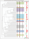
Figure 1
Figure 1
Phylogenetic analysis of the clonal and functional diversity of anti-EGFR antibodies. Cladogram showing the clonal and functional relationship between unique V gene rearrangements (clusters). The 24 selected antibodies that were analyzed in detail in this paper are highlighted in red, and genetically related antibody families or clusters originating from same V gene rearrangements are highlighted in color bars. Growth Inhibition HN5: Percentage growth inhibitory effect of antibody supernatants when tested by WST-1 assay on HN5 cells and compared with a negative control antibody. Negative values (green bars) indicate growth stimulatory effects.
Figure 2
Figure 2
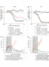
Functional evaluation of antibody mixtures. (A) Dose-response curves for inhibition of growth of the two cancer cell lines A431NS and HN5 by cetuximab. Cells were treated with cetuximab for 96 hours. The metabolic activity was measured by WST-1 addition and calculated as percentage of untreated control shown as mean ± SD. (B) Dose-response curves for inhibition of growth of the cancer cell line HN5 by 10 selected anti-EGFR antibodies and cetuximab after 96 hours of treatment with metabolic activity calculated as percentage of untreated control. (C) HN5 and A431NS cells treated with 2 µg/ml of the indicated antibody mixtures or cetuximab for 96 hours. The metabolic activity was measured by WST-1 addition and calculated as percentage of untreated control.
Figure 3
Figure 3
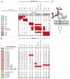
Epitope characterization of the selected set of 10 anti-EGFR antibodies. (A) Epitope binning by surface plasmon resonance competition assay. The anti-EGFR antibodies identified were binned according to EGFR domain by competition with murine reference antibodies with known EGFR specificity (data not shown) and according to cross-blocking. At least 50% inhibition (red) or 50% enhancement (green) were set as arbitrary cut-off values for antibody pairs binding overlapping epitopes or displaying binding co-operativity. (B) Mutational analysis of antibodies binding to EGFR variants detected by surface plasmon resonance analysis. The effect of binding to EGFR mutants is calculated as percentage maximum response level relative to wild type. Red color indicates less than 50% binding response compared with wild type. (C) Schematic diagram of the EGFR with the approximate location of the three domain III epitope bins shown.
Figure 4
Figure 4
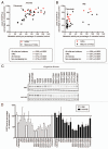
Correlation between level of growth inhibition and level of EGFR degradation. Plots derived from dot blot analysis showing the correlation between level of viable cells and level of EGFR degradation for mAbs and mixtures in (A) the A431NS cell line and (B) the HN5 cell line. (C) Immunoblots of cell lysates of HN5 or A431NS cells treated with mAbs or mixtures for 48 hours. (D) Odyssey quantification of band intensities from the immunoblots. EGFR levels were calculated as percentage of untreated control by using MFI values normalized to β-actin levels.
Figure 5
Figure 5
Functional evaluation of lead candidate mixtures in vitro and in vivo. Dose-response curves for inhibition of growth of the cell line A431NS (A) and HN5 (B) by the indicated antibodies and antibody mixtures. Cells were treated with antibody for 96 hours. The metabolic activity was measured by WST-1 addition and calculated as percentage of untreated control shown as mean ± SD. (C) The growth inhibitory effects of two lead candidate mixtures and cetuximab in A431NS human tumor xenografts. (D) The growth inhibitory effects of Sym004, the two individual antibodies 992 and 1024, and cetuximab in A431NS human tumor xenografts.

References

    1. Bublil EM, Yarden Y. The EGF receptor family: spearheading a merger of signaling and therapeutics. Curr Opin Cell Biol. 2007;19:124–134. - PubMed
    1. Harris RC, Chung E, Coffey RJ. EGF receptor ligands. Exp Cell Res. 2003;284:2–13. - PubMed
    1. Ogiso H, Ishitani R, Nureki O, Fukai S, Yamanaka M, Kim JH, et al. Crystal structure of the complex of human epidermal growth factor and receptor extracellular domains. Cell. 2002;110:775–787. - PubMed
    1. Garrett TP, McKern NM, Lou M, Elleman TC, Adams TE, Lovrecz GO, et al. Crystal structure of a truncated epidermal growth factor receptor extracellular domain bound to transforming growth factor alpha. Cell. 2002;110:763–773. - PubMed
    1. Ferguson KM, Berger MB, Mendrola JM, Cho HS, Leahy DJ, Lemmon MA. EGF activates its receptor by removing interactions that autoinhibit ectodomain dimerization. Mol Cell. 2003;11:507–517. - PubMed

MeSH terms