

Biochemical disclosure of the mycolate outer membrane of Corynebacterium glutamicum
- PMID: 22123248
- PMCID: PMC3264076
- DOI: 10.1128/JB.06138-11
Biochemical disclosure of the mycolate outer membrane of Corynebacterium glutamicum
Abstract
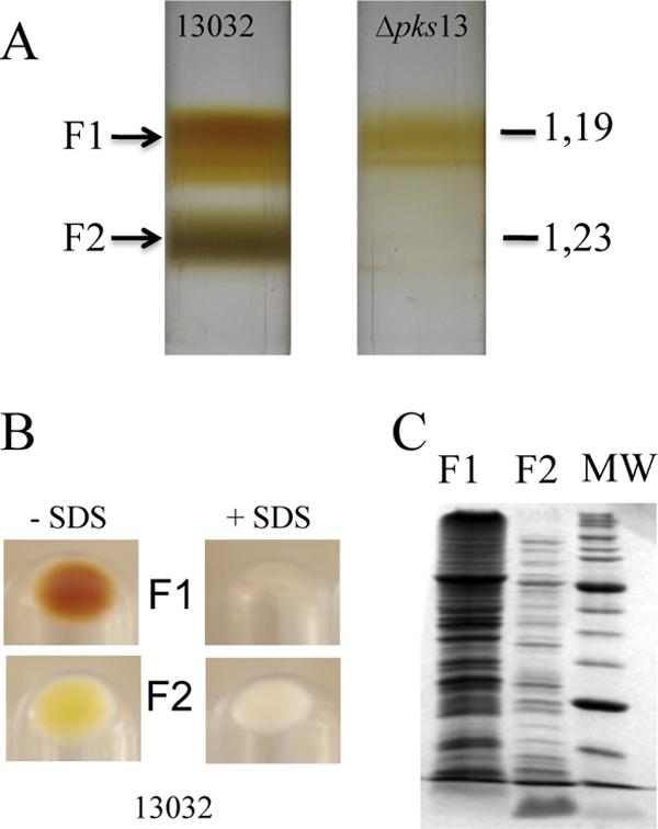
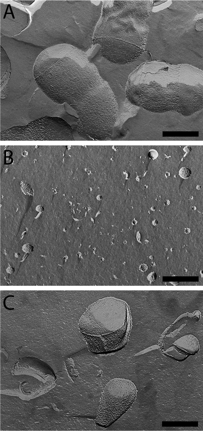
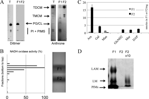
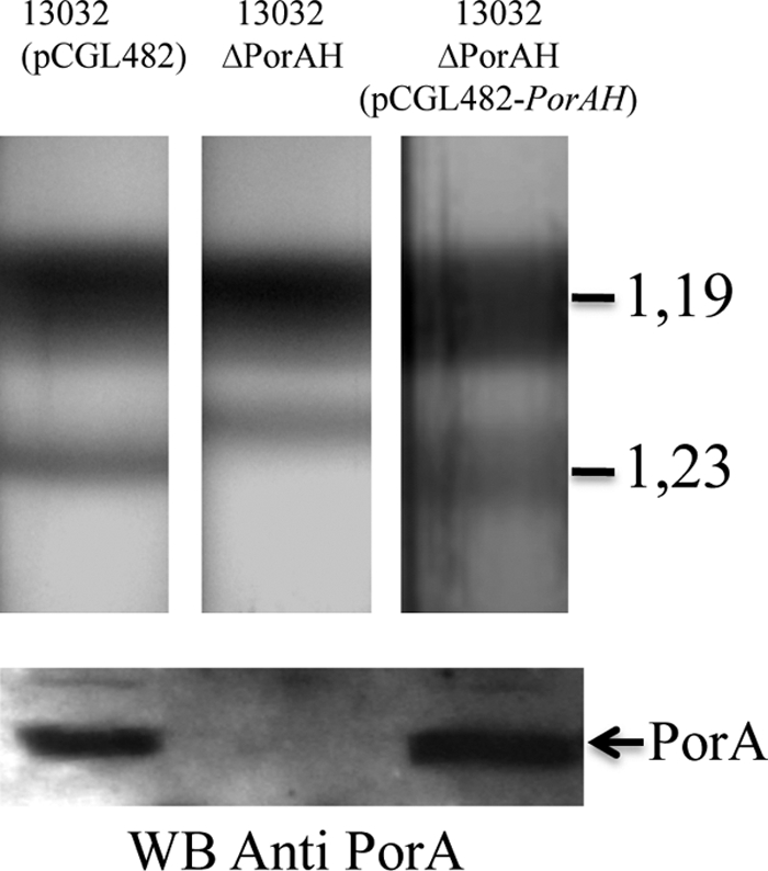
Corynebacterineae is a specific suborder of Gram-positive bacteria that includes Mycobacterium tuberculosis and Corynebacterium glutamicum. The cell wall of these bacteria is composed of a heteropolymer of peptidoglycan (PG) linked to arabinogalactan (AG), which in turn is covalently associated with an atypical outer membrane, here called mycomembrane (M). The latter structure has been visualized by cryo-electron microscopy of vitreous sections, but its biochemical composition is still poorly defined, thereby hampering the elucidation of its physiological function. In this report, we show for the first time that the mycomembrane-linked heteropolymer of PG and AG (M-AG-PG) of C. glutamicum can be physically separated from the inner membrane on a flotation density gradient. Analysis of purified M-AG-PG showed that the lipids that composed the mycomembrane consisted almost exclusively of mycolic acid derivatives, with only a tiny amount, if any, of phospholipids and lipomannans, which were found with the characteristic lipoarabinomannans in the plasma membrane. Proteins associated with or inserted in the mycomembrane were extracted from M-AG-PG with lauryl-dimethylamine-oxide (LDAO), loaded on an SDS-PAGE gel, and analyzed by tandem mass spectrometry or by Western blotting. Sixty-eight different proteins were identified, 19 of which were also found in mycomembrane fragments released by the terminal-arabinosyl-transferase-defective ΔAftB strain. Almost all of them are predicted to contain a signal sequence and to adopt the characteristic β-barrel structure of Gram-negative outer membrane proteins. These presumed mycomembrane proteins include the already-known pore-forming proteins (PorA and PorB), 5 mycoloyltransferases (cMytA, cMytB, cMytC, cMytD, and cMytF), several lipoproteins, and unknown proteins typified by a putative C-terminal hydrophobic anchor.
Figures
Similar articles
-
Identification of specific posttranslational O-mycoloylations mediating protein targeting to the mycomembrane.Proc Natl Acad Sci U S A. 2017 Apr 18;114(16):4231-4236. doi: 10.1073/pnas.1617888114. Epub 2017 Apr 3. Proc Natl Acad Sci U S A. 2017. PMID: 28373551 Free PMC article.
-
A deficiency in arabinogalactan biosynthesis affects Corynebacterium glutamicum mycolate outer membrane stability.J Bacteriol. 2010 Jun;192(11):2691-700. doi: 10.1128/JB.00009-10. Epub 2010 Apr 2. J Bacteriol. 2010. PMID: 20363942 Free PMC article.
-
Sequential assembly of the septal cell envelope prior to V snapping in Corynebacterium glutamicum.Nat Chem Biol. 2019 Mar;15(3):221-231. doi: 10.1038/s41589-018-0206-1. Epub 2019 Jan 21. Nat Chem Biol. 2019. PMID: 30664686 Free PMC article.
-
Current knowledge on mycolic acids in Corynebacterium glutamicum and their relevance for biotechnological processes.Appl Microbiol Biotechnol. 2013 Dec;97(23):9923-30. doi: 10.1007/s00253-013-5265-3. Epub 2013 Oct 11. Appl Microbiol Biotechnol. 2013. PMID: 24113823 Review.
-
Mycomembrane and S-layer: two important structures of Corynebacterium glutamicum cell envelope with promising biotechnology applications.J Biotechnol. 2003 Sep 4;104(1-3):55-67. doi: 10.1016/s0168-1656(03)00163-9. J Biotechnol. 2003. PMID: 12948629 Review.
Cited by
-
Cell envelope of corynebacteria: structure and influence on pathogenicity.ISRN Microbiol. 2013 Jan 21;2013:935736. doi: 10.1155/2013/935736. Print 2013. ISRN Microbiol. 2013. PMID: 23724339 Free PMC article.
-
A chromosomally encoded T7 RNA polymerase-dependent gene expression system for Corynebacterium glutamicum: construction and comparative evaluation at the single-cell level.Microb Biotechnol. 2015 Mar;8(2):253-65. doi: 10.1111/1751-7915.12236. Epub 2014 Dec 9. Microb Biotechnol. 2015. PMID: 25488698 Free PMC article.
-
Identification of specific posttranslational O-mycoloylations mediating protein targeting to the mycomembrane.Proc Natl Acad Sci U S A. 2017 Apr 18;114(16):4231-4236. doi: 10.1073/pnas.1617888114. Epub 2017 Apr 3. Proc Natl Acad Sci U S A. 2017. PMID: 28373551 Free PMC article.
-
Native presynaptic metabotropic glutamate receptor 4 (mGluR4) interacts with exocytosis proteins in rat cerebellum.J Biol Chem. 2012 Jun 8;287(24):20176-86. doi: 10.1074/jbc.M112.347468. Epub 2012 Apr 23. J Biol Chem. 2012. PMID: 22528491 Free PMC article.
-
Complete genome sequence of Corynebacterium pseudotuberculosis biovar ovis strain P54B96 isolated from antelope in South Africa obtained by rapid next generation sequencing technology.Stand Genomic Sci. 2012 Dec 19;7(2):189-99. doi: 10.4056/sigs.3066455. Epub 2012 Dec 15. Stand Genomic Sci. 2012. PMID: 23408795 Free PMC article.
References
-
- Aggerbeck LP, Gulik-Krzywicki T. 1986. Studies of lipoproteins by freeze-fracture and etching electron microscopy. Methods Enzymol. 128:457–472 - PubMed
Publication types
MeSH terms
Substances
LinkOut - more resources
Full Text Sources
Molecular Biology Databases
