Review
. 2011 Nov 26;11(1):166-74.
doi: 10.1102/1470-7330.2011.0023.

Extranodal manifestations of lymphoma on [¹⁸F]FDG-PET/CT: a pictorial essay

Affiliations
Review

Extranodal manifestations of lymphoma on [¹⁸F]FDG-PET/CT: a pictorial essay

Raghava Kashyap et al. Cancer Imaging. .

Abstract

Lymphoma is the seventh most common type of malignancy in both sexes. It is a neoplastic proliferation of lymphoid cells at various stages of differentiation and affects lymph nodes with infiltration into the bone marrow, spleen and thymus. However, extra nodal involvement is frequently seen in many cases. With the development of dedicated positron emission tomography (PET) scanners with fused computed tomographic (CT) systems in the same gantry, [18F]fluorodeoxyglucose (FDG)-PET/CT has become a major tool in the evaluation of lymphomas and it is inimitable in certain situations such as assessment of response to therapy. Extranodal lymphoma can present with diverse manifestations and sometimes mimics other organ-related pathologies. Knowledge of the protean manifestations of extranodal lymphoma is required to accurately detect the disease and differentiate it from the various physiologic and benign causes of FDG uptake in various organs. We present a case series of extranodal involvement of histologically proven cases of lymphomas detected on FDG-PET/CT at our institute to demonstrate the challenges in interpretation of extranodal lymphoma.


Figures

Figure 1
Figure 1
A 43-year-old man with a history of seizures. PET scan showed a hypermetabolic lesion in the cerebellum (A,D) which after craniotomy (B,E) and biopsy turned out to be primary CNS lymphoma involving the cerebellar region of diffuse large B cell subtype. Chemotherapy with methotrexate showed good response (C,F). PET scan in this case helped rule out demyelination, which was one of the differentials at the presentation, by showing the lesion to be intensely hypermetabolic.
Figure 2
Figure 2
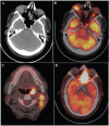
(A) CT of a 46-year-old man showing a homogeneous soft tissue lesion in the right orbit. (B) Intense FDG uptake is seen in the orbital lesion, which was proven to be diffuse large B cell lymphoma. (C) Tonsillar lymphoma in a 53-year-old man of T cell subtype. (D) Lymphoma of T cell subtype involving the ethmoid sinus on the left side in a 62-year-old male patient. All these lesions were found on PET/CT scans that were referred for staging purposes.
Figure 3
Figure 3
Intense FDG uptake in a destructive soft tissue lesion involving the nasal septum in a case of NK/T cell lymphoma, also called midline lethal granuloma/ulcerating midline granuloma, in a 38-year-old man, which was found on a staging PET/CT scan. The patient had presented with foul smelling nasal discharge, which led to the diagnosis of lymphoma.
Figure 4
Figure 4
Various manifestations of lymphoma involving the lungs. (A) Nodular presentation of diffuse large B cell lymphoma in a 39-year-old man. (B) Hodgkin disease presenting as a mass lesion in a 24-year-old man. (C,D) NHL presenting as a mass-like consolidation in the right lung with intense uptake along with pleural effusion on the right side in a 28-year-old woman.
Figure 5
Figure 5
T cell NHL in a 35-year-old man presenting as multiple large mass-like deposits (arrows) along the pleura associated with large pleural effusion (*) on the left side.
Figure 6
Figure 6
A 68-year-old woman with bilateral breast lumps, which on PET/CT showed intense FDG avidity. The lesions were histologically proven to be diffuse large B cell lymphoma.
Figure 7
Figure 7
NHL showing intense FDG uptake in the anteropyloric region of the stomach along with perigastric lymph nodes in a 70-year-old man. Gastric lymphomas are usually associated with H. pylori gastritis.
Figure 8
Figure 8
A 30-year-old male renal transplant patient (which is visible in the right iliac region). Intense FDG uptake is seen along the grossly thickened wall of a large segment of small bowel. This was confirmed to be plasmablastic variant of B cell lymphoma.
Figure 9
Figure 9
PET/CT of a 70-year-old woman presenting with pyrexia of unknown origin (PUO) showed intense diffuse uptake in the liver and multiple foci in the bone marrow that was confirmed as NHL of the liver and was CD 20 positive. PET/CT was used for the investigation in this case to evaluate the PUO persisting for 2 months and the patient showed lesions only at extranodal sites, including the bone marrow, apart from the liver.
Figure 10
Figure 10
A 66-year-old woman presenting with a flank mass on right side, which shows intense FDG uptake within a large homogeneous lesion in the right kidney along with renal hilar nodes on PET/CT. This was proven to be NHL. Primary renal neoplasms usually present as heterogeneously enhancing masses with low to variable uptake on FDG. Therefore in this case, PET/CT prompted the clinicians to undertake fine-needle aspiration cytology, which otherwise is not preferred for suspicion of renal cell carcinoma thereby helping in the diagnosis.
Figure 11
Figure 11
Lymphoma of T cell type involving both the adrenal glands in 62-year-old man. Bilateral involvement is seen in up to 50% cases in which the adrenals are involved.
Figure 12
Figure 12
Intense asymmetric uptake in the grossly enlarged left testis in a 62-year-old man. The patient was diagnosed with B cell NHL on the right side for which he was subjected to orchidectomy followed by chemotherapy. He presented with enlargement of the left side, which was proven to be NHL.
Figure 13
Figure 13
B cell NHL involving the body of the uterus in a 53-year-old woman.
Figure 14
Figure 14
T cell cutaneous lymphoma showing intense FDG uptake in a skin lesion and subcutaneous nodules in a 62-year-old man.
Figure 15
Figure 15
Diffuse large cell lymphoma involving the vertebra and adjacent rib causing expansile lytic destruction in a 40-year-old woman.

References

    1. Altekruse SF, Kosary CL, Krapcho M, et al. SEER Cancer statistics review, 1975–2007. National Cancer Institute, Bethesda, MD. http://seer.cancer.gov/csr/1975_2007/, based on November 2009 SEER data submission, posted to the SEER web site, 2010.
    1. Freeman C, Berg JW, Cutler SJ. Occurrence and prognosis of extranodal lymphomas. Cancer. 1972;29:252–60. doi: 10.1002/1097-0142(197201)29:1<252::AID-CNCR2820290138>3.0.CO;2-#. - DOI - PubMed
    1. Glass AG, Karnell LH, Menck HR. The National Cancer Data Base report on non-Hodgkin's lymphoma. Cancer. 1997;80:2311–20. doi: 10.1002/(SICI)1097-0142(19971215)80:12<2311::AID-CNCR13>3.3.CO;2-U. - DOI - PubMed
    1. Guermazi A, Brice P, de Kerviler EE, Fermé C, Hennequin C, Meignin V. Extranodal Hodgkin disease: spectrum of disease. Radiographics. 2001;21:161–79. - PubMed
    1. Paes FM, Kalkanis DG, Sideras PA, Serafini AN. FDG PET/CT of extranodal involvement in non-Hodgkin lymphoma and Hodgkin disease. Radiographics. 2010;30:269–91. doi: 10.1148/rg.301095088. - DOI - PubMed

MeSH terms

Substances