

Characterization of NOL7 gene point mutations, promoter methylation, and protein expression in cervical cancer
- PMID: 22123719
- PMCID: PMC3237951
- DOI: 10.1097/PGP.0b013e318220ba16
Characterization of NOL7 gene point mutations, promoter methylation, and protein expression in cervical cancer
Abstract
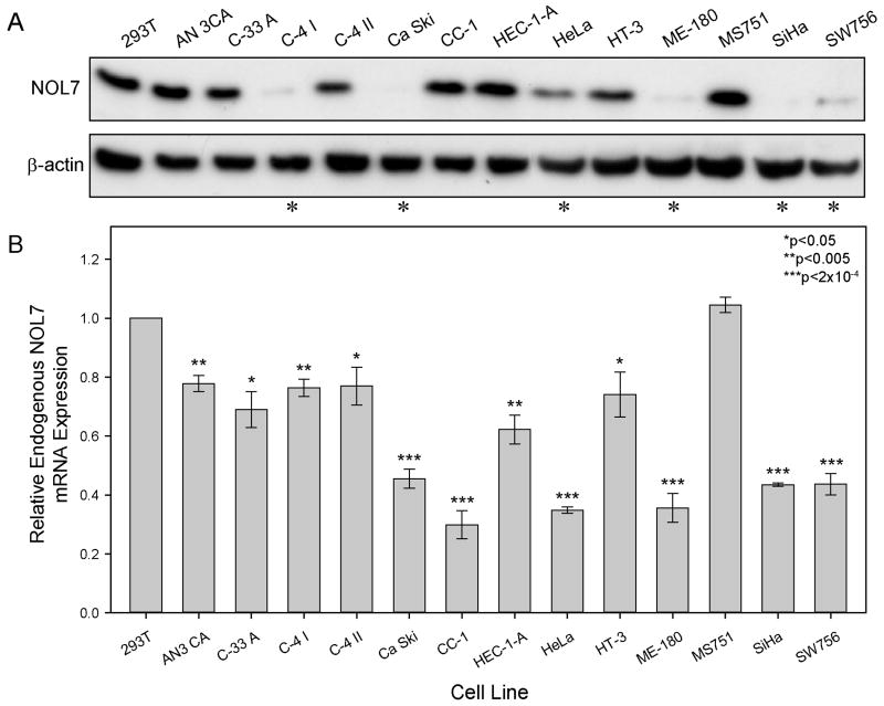
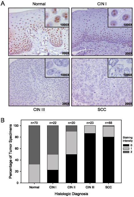
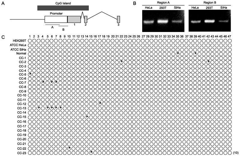
NOL7 is a putative tumor suppressor gene localized to 6p23, a region with frequent loss of heterozygosity in a number of cancers, including cervical cancer (CC). We have previously demonstrated that reintroduction of NOL7 into CC cells altered the angiogenic phenotype and suppressed tumor growth in vivo by 95%. Therefore, to understand its mechanism of inactivation in CC, we investigated the genetic and epigenetic regulation of NOL7. NOL7 mRNA and protein levels were assessed in 13 CC cell lines and 23 consecutive CC specimens by real-time quantitative polymerase chain reaction, western blotting, and immunohistochemistry. Methylation of the NOL7 promoter was analyzed by bisulfite sequencing and mutations were identified through direct sequencing. A CpG island with multiple CpG dinucleotides spanned the 5' untranslated region and first exon of NOL7. However, bisulfite sequencing failed to identify persistent sites of methylation. Mutational sequencing revealed that 40% of the CC specimens and 31% of the CC cell lines harbored somatic mutations that may affect the in vivo function of NOL7. Endogenous NOL7 mRNA and protein expression in CC cell lines were significantly decreased in 46% of the CC cell lines. Finally, immunohistochemistry demonstrated strong NOL7 nucleolar staining in normal tissues that decreased with histologic progression toward CC. NOL7 is inactivated in CC in accordance with the Knudson 2-hit hypothesis through loss of heterozygosity and mutation. Together with evidence of its in vivo tumor suppression, these data support the hypothesis that NOL7 is the legitimate tumor suppressor gene located on 6p23.
Conflict of interest statement
Figures
References
-
- Jemal A, Bray F, Center MM, Ferlay J, Ward E, Forman D. Global cancer statistics. CA Cancer J Clin. 2011 Mar-Apr;61(2):69–90. - PubMed
-
- Narisawa-Saito M, Yoshimatsu Y, Ohno S, et al. An in vitro multistep carcinogenesis model for human cervical cancer. Cancer Res. 2008 Jul 15;68(14):5699–5705. - PubMed
-
- Branca M, Giorgi C, Ciotti M, et al. Down-regulated nucleoside diphosphate kinase nm23-H1 expression is unrelated to high-risk human papillomavirus but associated with progression of cervical intraepithelial neoplasia and unfavourable prognosis in cervical cancer. J Clin Pathol. 2006 Oct;59(10):1044–1051. - PMC - PubMed
-
- Kaufmann AM, Backsch C, Schneider A, Durst M. HPV induced cervical carcinogenesis: molecular basis and vaccine development. Zentralbl Gynakol. 2002 Nov;124(11):511–524. - PubMed
-
- Walboomers JM, Jacobs MV, Manos MM, et al. Human papillomavirus is a necessary cause of invasive cervical cancer worldwide. J Pathol. 1999 Sep;189(1):12–19. - PubMed
Publication types
MeSH terms
Substances
Grants and funding
LinkOut - more resources
Full Text Sources
Medical
