Review
. 2011 Dec 12;195(6):931-42.
doi: 10.1083/jcb.201108081. Epub 2011 Nov 28.

Cell death and infection: a double-edged sword for host and pathogen survival

Affiliations
Review

Cell death and infection: a double-edged sword for host and pathogen survival

Hiroshi Ashida et al. J Cell Biol. .

Abstract

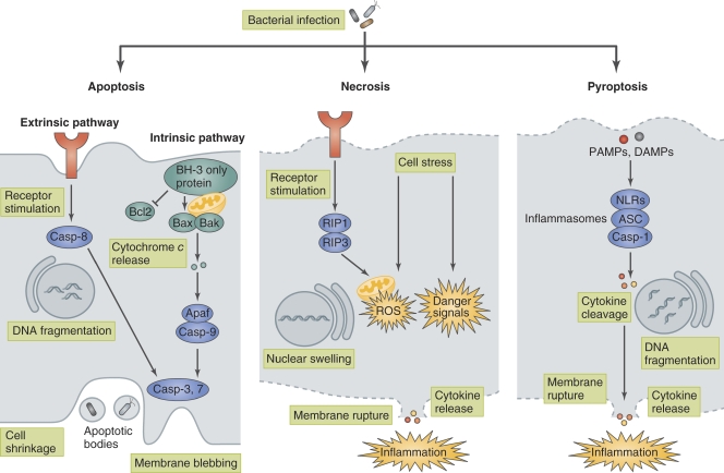
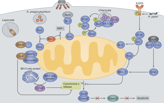
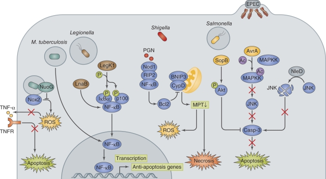
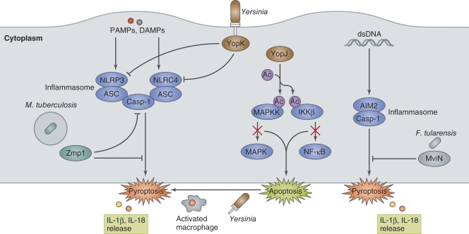
Host cell death is an intrinsic immune defense mechanism in response to microbial infection. However, bacterial pathogens use many strategies to manipulate the host cell death and survival pathways to enhance their replication and survival. This manipulation is quite intricate, with pathogens often suppressing cell death to allow replication and then promoting it for dissemination. Frequently, these effects are exerted through modulation of the mitochondrial pro-death, NF-κB-dependent pro-survival, and inflammasome-dependent host cell death pathways during infection. Understanding the molecular details by which bacterial pathogens manipulate cell death pathways will provide insight into new therapeutic approaches to control infection.


Figures

Figure 1.
Figure 1.
Bacteria-induced host cell death. Bacteria induce host cell death through several distinct modalities, including apoptosis, necrosis, and pyroptosis. Apoptosis is a type of noninflammatory programmed cell death that is triggered by two different pathways, the intrinsic (mitochondria-mediated) pathway and extrinsic (receptor-mediated) pathway. Apoptosis is morphologically characterized by membrane blebbing, cell shrinkage, DNA fragmentation, mitochondrial permeability, and caspase (except for caspase-1) activation. In apoptosis, bacteria are retained within apoptotic bodies and engulfed by phagocytic cells. Necrosis is characterized by membrane rupture, nuclear swelling, and the release of cellular contents and is accompanied by caspase-independent inflammation. Necrosis is triggered by ROS production or danger signals, such as lysosomal destabilization, calpain release, and depletion of ATP, that are induced upon bacterial infection or physical damage. Pyroptosis is a type of programmed cell death that is coordinated by inflammasome-mediated caspase-1 activation and accompanied by membrane rupture, DNA fragmentation, and the release of pro-inflammatory cytokines, including IL-1β and IL-18. PAMPs and DAMPs are recognized by NLR proteins, which assemble the inflammasome to activate caspase-1and trigger pyroptosis.
Figure 2.
Figure 2.
Bacterial manipulation of mitochondrial cell death pathways. Legionella deliver SidF and SdhA. SidF interacts with two pro-apoptotic proteins, BNIP3 and Bcl-rambo, and inhibits apoptotic signaling. SdhA inhibits host cell death and enhances bacterial survival and replication by an as yet unknown mechanism. A. phagocytophilum delivers Ats-1 and prevents the loss of mitochondrial membrane potential (MMP) and Bax translocation to the mitochondria to inhibit apoptosis. EPEC delivers NleH, which interacts with Bax inhibitor 1 (BI-1) and blocks epithelial apoptosis. Chlamydia secrete CPAF, which degrades the pro-apoptotic BH3-only proteins, thereby blocking the release of cytochrome c from the mitochondria. Chlamydia infection also inhibits the function of the pro-apoptotic protein Bad by causing its phosphorylation by Akt and mislocalization to Chlamydia inclusion vacuoles via its interaction with 14-3-3β and IncG. H. pylori delivers the T4SS effector CagA to attenuate epithelial cell apoptosis. The CagA and Grb2 or Crk interaction stimulates the ERK1/2 pathway and up-regulates the production of the anti-apoptotic factor MCL-1. H. pylori activates EGFR, thereby up-regulating anti-apoptotic signaling involving Akt and Bcl2.
Figure 3.
Figure 3.
Bacterial manipulation of host pro-survival pathway. M. tuberculosis activates host anti-apoptotic signaling by up-regulating the anti-apoptosis genes Mcl-1, bfl1, and FLIP. NuoG dampens Nox2-mediated host signaling, resulting in the inhibition of apoptosis. Legionella deliver LegK1 and LnaB, which enhance the pro-survival activity of NF-κB. Shigella invasion of the epithelium results in mitochondria permeability transition (MPT)−dependent ROS production and necrosis-like cell death, which is counterbalanced by the NOD1−RIP2−NF-κB−Bcl2 pro-survival pathway. PGN, peptidoglycan. Salmonella deliver SopB and AvrA via the T3SS, and they inhibit apoptosis by activating the PI3K–Akt pro-survival pathway via inositol phosphate activity and by preventing JNK pro-cell death signaling via acetyltransferase activity, respectively. EPEC also targets JNK signaling by delivering NleD, which uses its metalloprotease activity to cleave JNK, thereby inhibiting cell death.
Figure 4.
Figure 4.
Bacterial manipulation of inflammasome activation. PAMPs or DAMPs (such as dsDNA) generated by bacterial invasion and multiplication in macrophages trigger NLRP3-, NLRC4-, AIM2-inflammasome assembly and induce pyroptosis. Yersinia induce apoptosis by YopJ-mediated inhibition of pro-inflammatory signaling in the initial stage of infection in naive macrophages. Activated macrophages redirect YopJ-mediated apoptosis to YopJ-independent pyroptosis. However, Yersinia prevent inflammasome activation by secreting YopK, which interferes with T3SS recognition by NLRP3 and NLRC4. M. tuberculosis Zmp1, a Zn metalloprotease, and F. tularensis MviN, a putative lipid II flippase, prevent inflammasome activation and IL-1β secretion/pyroptosis by unknown mechanisms.

References

    1. Abdul-Sater A.A., Koo E., Häcker G., Ojcius D.M. 2009. Inflammasome-dependent caspase-1 activation in cervical epithelial cells stimulates growth of the intracellular pathogen Chlamydia trachomatis. J. Biol. Chem. 284:26789–26796 10.1074/jbc.M109.026823 - DOI - PMC - PubMed
    1. Abu-Zant A., Jones S., Asare R., Suttles J., Price C., Graham J., Kwaik Y.A. 2007. Anti-apoptotic signalling by the Dot/Icm secretion system of L. pneumophila. Cell. Microbiol. 9:246–264 10.1111/j.1462-5822.2006.00785.x - DOI - PubMed
    1. Amer A., Franchi L., Kanneganti T.D., Body-Malapel M., Ozören N., Brady G., Meshinchi S., Jagirdar R., Gewirtz A., Akira S., Núñez G. 2006. Regulation of Legionella phagosome maturation and infection through flagellin and host Ipaf. J. Biol. Chem. 281:35217–35223 10.1074/jbc.M604933200 - DOI - PubMed
    1. Amieva M.R., El-Omar E.M. 2008. Host-bacterial interactions in Helicobacter pylori infection. Gastroenterology. 134:306–323 10.1053/j.gastro.2007.11.009 - DOI - PubMed
    1. Arnoult D., Carneiro L., Tattoli I., Girardin S.E. 2009. The role of mitochondria in cellular defense against microbial infection. Semin. Immunol. 21:223–232 10.1016/j.smim.2009.05.009 - DOI - PubMed

Publication types