Review
. 2011 Dec 26;195(7):1071-82.
doi: 10.1083/jcb.201108131. Epub 2011 Nov 28.

The cell biology of receptor-mediated virus entry

Affiliations
Review

The cell biology of receptor-mediated virus entry

Joe Grove et al. J Cell Biol. .

Abstract

The cell imposes multiple barriers to virus entry. However, viruses exploit fundamental cellular processes to gain entry to cells and deliver their genetic cargo. Virus entry pathways are largely defined by the interactions between virus particles and their receptors at the cell surface. These interactions determine the mechanisms of virus attachment, uptake, intracellular trafficking, and, ultimately, penetration to the cytosol. Elucidating the complex interplay between viruses and their receptors is necessary for a full understanding of how these remarkable agents invade their cellular hosts.


Figures

Figure 1.
Figure 1.
Virus entry strategies. The cell imposes intrinsic barriers to virus entry including the plasma membrane, actin cortex, and limiting intracellular membranes. (A and B) Viruses have evolved various strategies to overcome these barriers, such as receptor-mediated endocytosis followed by pH-dependent/independent fusion from endocytic compartments (A) or pH-independent fusion at the plasma membrane, coupled with receptor-mediated signaling and coordinated disassembly of the actin cortex (B). Enveloped viruses are shown; nonenveloped viruses use similar strategies, although the mechanisms of action are different. MVB, multivesicular body.
Figure 2.
Figure 2.
Virus receptors. Virus entry is initiated by specific interactions between virus particles and receptors. (A) Human rhinovirus 2 undergoes receptor-mediated endocytosis after interaction with LDLR. (B) CD4 is the primary receptor for HIVs, but virus penetration requires further interactions with chemokine receptor CCR5 or CXCR4. Initial observations indicated that coreceptor engagement triggered fusion directly at the plasma membrane; however, recent studies suggest that fusion can also occur after endocytosis (Miyauchi et al., 2009). Although these mechanisms appear mutually exclusive, it is possible that both may operate, and additional studies are required to establish the relevant pathway for key target cells in vivo. (C) HCV entry requires at least four host factors. The virus particle is thought to directly interact with SR-B1 and CD81, whereas the tight junction components claudin-1 and occludin are indirectly involved. Data suggest that CD81/claudin-1 heteromers are necessary for infection. It is currently unknown how HCV is directed to clathrin-coated vesicles.
Figure 3.
Figure 3.
Sites of virus particle fusion/penetration. Virus particles must transport genetic material across limiting membranes; this can be achieved at various locations within the cell. (A) Enveloped virus particles can fuse directly at the plasma membrane at neutral pH after interaction with cell surface receptors. (B and C) Alternatively, internalized virus particles can escape from the endosomal network. This is often dependent on endosome acidification and occurs at either mild pH (6.5–6) from the early endosome (B) or low pH (5.5–4) from late endosome and/or lysosome (C). In addition to the acidic environment, other molecular cues may be required to trigger fusion/penetration, for example, membrane lipid content (Semliki Forest virus and Dengue virus) or proteolytic cleavage (reovirus and SARS coronavirus; Skehel et al., 1982; Schlegel and Wade, 1984; Mothes et al., 2000; Brabec et al., 2003). (D) Polyomaviruses such as SV40 undergo atypical transport through the endosomal pathway to the ER, where partially disassembled virus particles are shuttled to the cytosol by the retrotranslocation machinery.
Figure 4.
Figure 4.
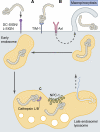
Ebola virus entry. (A) Lectins DC-SIGN and L-SIGN act as attachment factors to concentrate Ebola virus particles at the cell surface (Alvarez et al., 2002; Simmons et al., 2003), facilitating interaction with the receptor TIM-1. (B) Axl receptor tyrosine kinase is thought to promote virus particle uptake via macropinocytosis. Critically, Ebola virus does not directly engage Axl; the Axl ligand Gas-6 may associate with virus particles and facilitate indirect interaction between Ebola virus and Axl, as demonstrated for other viruses (Morizono et al., 2011). (C) Within the late endosome/lysosome, viral glycoprotein GP1 undergoes sequential proteolytic cleavage by cathepsins L and B, allowing interaction with NPC1, a putative endosomal receptor. Ebola virus membrane fusion is dependent on the viral glycoprotein GP2 and occurs from the late endosome/lysosome, although the exact molecular triggers remain unclear.

References

    1. Alvarez C.P., Lasala F., Carrillo J., Muñiz O., Corbí A.L., Delgado R. 2002. C-type lectins DC-SIGN and L-SIGN mediate cellular entry by Ebola virus in cis and in trans. J. Virol. 76:6841–6844 10.1128/JVI.76.13.6841-6844.2002 - DOI - PMC - PubMed
    1. Amstutz B., Gastaldelli M., Kälin S., Imelli N., Boucke K., Wandeler E., Mercer J., Hemmi S., Greber U.F. 2008. Subversion of CtBP1-controlled macropinocytosis by human adenovirus serotype 3. EMBO J. 27:956–969 10.1038/emboj.2008.38 - DOI - PMC - PubMed
    1. Barth H., Schafer C., Adah M.I., Zhang F., Linhardt R.J., Toyoda H., Kinoshita-Toyoda A., Toida T., Van Kuppevelt T.H., Depla E., et al. 2003. Cellular binding of hepatitis C virus envelope glycoprotein E2 requires cell surface heparan sulfate. J. Biol. Chem. 278:41003–41012 10.1074/jbc.M302267200 - DOI - PubMed
    1. Barton E.S., Forrest J.C., Connolly J.L., Chappell J.D., Liu Y., Schnell F.J., Nusrat A., Parkos C.A., Dermody T.S. 2001. Junction adhesion molecule is a receptor for reovirus. Cell. 104:441–451 10.1016/S0092-8674(01)00231-8 - DOI - PubMed
    1. Bergelson J.M., Cunningham J.A., Droguett G., Kurt-Jones E.A., Krithivas A., Hong J.S., Horwitz M.S., Crowell R.L., Finberg R.W. 1997. Isolation of a common receptor for Coxsackie B viruses and adenoviruses 2 and 5. Science. 275:1320–1323 10.1126/science.275.5304.1320 - DOI - PubMed

Publication types

Substances