Review
. 2011 Nov 28;195(5):709-20.
doi: 10.1083/jcb.201102131.

Cell cycle regulation in hematopoietic stem cells

Affiliations
Review

Cell cycle regulation in hematopoietic stem cells

Eric M Pietras et al. J Cell Biol. .

Abstract

Hematopoietic stem cells (HSCs) give rise to all lineages of blood cells. Because HSCs must persist for a lifetime, the balance between their proliferation and quiescence is carefully regulated to ensure blood homeostasis while limiting cellular damage. Cell cycle regulation therefore plays a critical role in controlling HSC function during both fetal life and in the adult. The cell cycle activity of HSCs is carefully modulated by a complex interplay between cell-intrinsic mechanisms and cell-extrinsic factors produced by the microenvironment. This fine-tuned regulatory network may become altered with age, leading to aberrant HSC cell cycle regulation, degraded HSC function, and hematological malignancy.


Figures

Figure 1.
Figure 1.
Distinct cell cycle activity in fetal, adult, and old HSCs. HSC cell cycle activity and cellular output is highly dynamic throughout the lifetime of an organism. During fetal life (left column), HSCs display a high level of cell cycle activity, as their primary role at this stage is the genesis of the nascent blood system. During adult life (center column), HSCs reside in the BM and enter a predominantly quiescent (G0) state, generating a balanced myeloid, erythroid, and lymphoid output to maintain blood homeostasis. As individuals age (right column), HSCs appear to remain predominantly quiescent, but their function begins to degrade (dotted outlines), resulting in the loss of erythroid and lymphoid output and bias toward the myeloid lineage. AGM: aorta-gonad-mesonephros; ND: not determined. See text for detailed discussion and references.
Figure 2.
Figure 2.
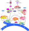
HSC cell cycle entry is regulated by a complex network of cell-intrinsic and cell-extrinsic factors. The entry of quiescent HSCs from G0 into the G1 phase of the cell cycle is governed primarily via competing activating and inhibitory mechanisms that regulate the activity of cyclin–Cdk complexes. The PI3K/Akt/mTOR pathway, which is activated in response to numerous extrinsic signals, is considered a central activator of HSC cell cycle activity, primarily via activation of the cyclin D–Cdk4/6 complex. This pathway is heavily regulated, primarily by PTEN and TSC1/2. Moreover, the Ink4 CKI family inhibits cyclin D–Cdk4/6 activity, and CIP/KIP family CKIs are also capable of inhibiting Cdk4 activity. Progression from the G1 to the S phase of the cell cycle is regulated by Cyclin E–Cdk2. This complex is regulated via the CIP/KIP family of Cdk inhibitors, as well as by the Rb family. Expression of CIP/KIP family members is in turn regulated by transcription factors such as Hes1, JunB, and FoxO3a, which are activated by extrinsic growth-repressive signals. Furthermore, HSC cell cycle activity is subject to regulation via p53, either in response to cellular damage or p19ARF activity. Solid arrows indicate direct activation/inhibition events, dashed arrows indicate transcriptional regulation events. Functionally related groups of cell cycle activators are shaded in green; functionally related groups of cell cycle inhibitors are shaded in red.

References

    1. Adams G.B., Chabner K.T., Alley I.R., Olson D.P., Szczepiorkowski Z.M., Poznansky M.C., Kos C.H., Pollak M.R., Brown E.M., Scadden D.T. 2006. Stem cell engraftment at the endosteal niche is specified by the calcium-sensing receptor. Nature. 439:599–603 10.1038/nature04247 - DOI - PubMed
    1. Arai F., Hirao A., Ohmura M., Sato H., Matsuoka S., Takubo K., Ito K., Koh G.Y., Suda T. 2004. Tie2/angiopoietin-1 signaling regulates hematopoietic stem cell quiescence in the bone marrow niche. Cell. 118:149–161 10.1016/j.cell.2004.07.004 - DOI - PubMed
    1. Asai T., Liu Y., Bae N., Nimer S.D. 2011. The p53 tumor suppressor protein regulates hematopoietic stem cell fate. J. Cell. Physiol. 226:2215–2221 10.1002/jcp.22561 - DOI - PMC - PubMed
    1. Attema J.L., Pronk C.J.H., Norddahl G.L., Nygren J.M., Bryder D. 2009. Hematopoietic stem cell ageing is uncoupled from p16 INK4A-mediated senescence. Oncogene. 28:2238–2243 10.1038/onc.2009.94 - DOI - PubMed
    1. Baldridge M.T., King K.Y., Boles N.C., Weksberg D.C., Goodell M.A. 2010. Quiescent haematopoietic stem cells are activated by IFN-gamma in response to chronic infection. Nature. 465:793–797 10.1038/nature09135 - DOI - PMC - PubMed

Substances