

Screening for carpal tunnel syndrome using sonography
- PMID: 22124001
- PMCID: PMC3654536
- DOI: 10.7863/jum.2011.30.12.1657
Screening for carpal tunnel syndrome using sonography
Abstract
Objectives: The use of sonography in musculoskeletal research and clinical applications is increasing; however, measurement techniques for diagnosing carpal tunnel syndrome with sonography continue to be inconsistent. Novel methods of measurement using internal comparisons to identify swelling of the median nerve require investigation and comparison to currently used techniques.
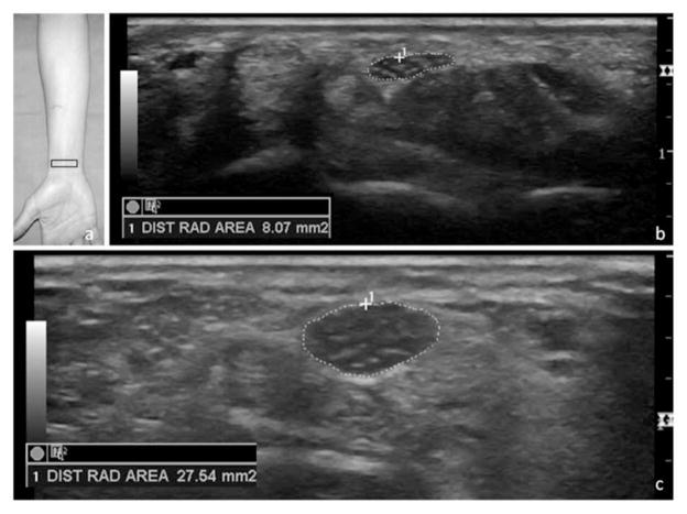
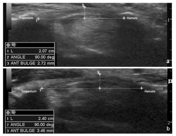
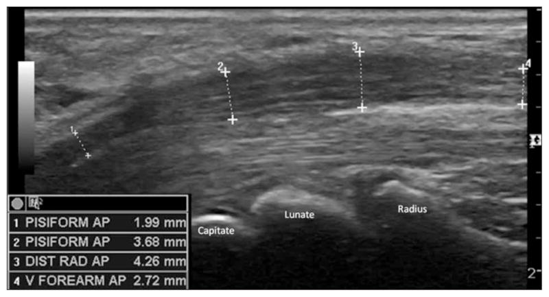
Methods: The flattening ratio of the median nerve, bowing of the flexor retinaculum, and cross-sectional area of the median nerve were collected in the forearm, at the radiocarpal joint, and at the level of the pisiform in both symptomatic patients and asymptomatic control participants. Electrodiagnostic testing was completed in symptomatic patients as a diagnostic standard.
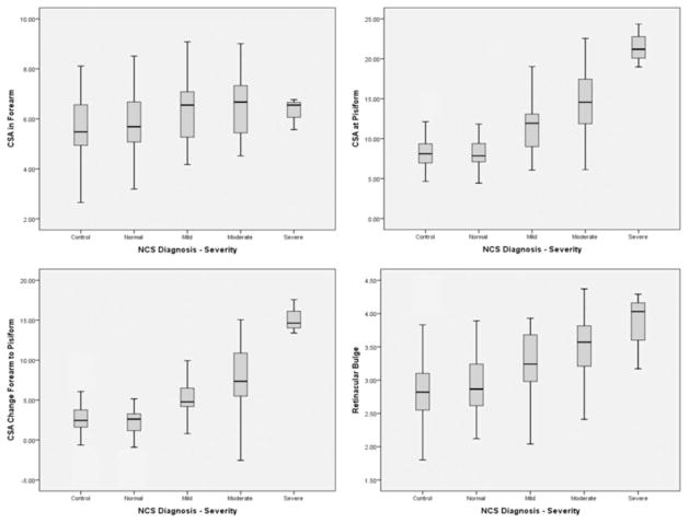
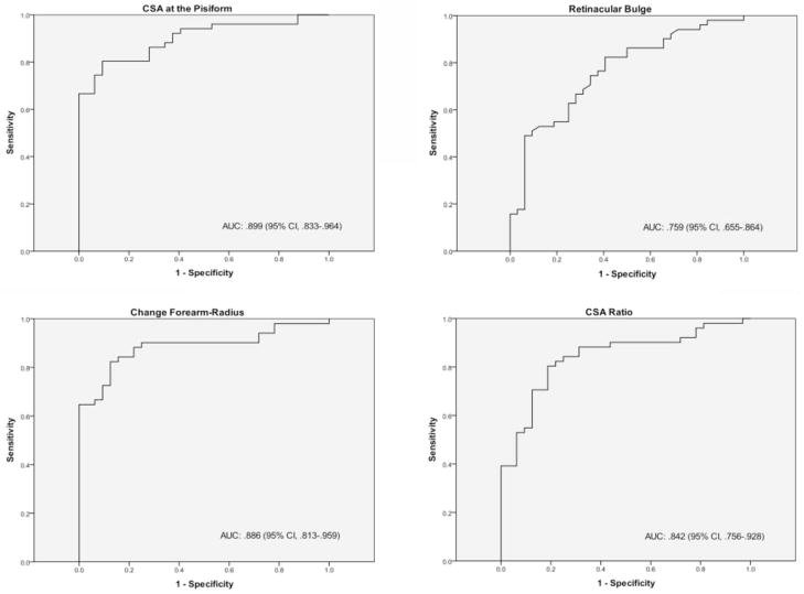
Results: Median nerve measurements were collected from 166 wrists of symptomatic and asymptomatic participants. The flattening ratio did not show any correlation to electrodiagnostic testing and was identical between both symptomatic and asymptomatic participants. Moderate to strong correlations were noted between electrodiagnostic testing results and sonographic measurements of the cross-sectional area at the pisiform, retinacular bowing, and both the ratio and change of the cross-sectional area between the forearm and pisiform. The area under the curve was large for all receiver operating characteristic curves for each measurement (0.759-0.899), and sensitivity was high (80.4%-82.4%).
Conclusions: Measurement of swelling through a ratio or absolute change had similar diagnostic accuracy as individual measurement of the cross-sectional area within the carpal tunnel. These measures may be useful for improving accuracy in more diverse clinical populations. Further refinement of protocols to identify the largest cross-sectional area within the carpal tunnel region and statistical methods to analyze clustered, multilevel outcome data are recommended to improve diagnostics.
Figures
References
-
- Roll SC, Evans K. Feasibility of using a hand-carried sonographic unit for investigating median nerve pathology. J Diagn Med Sonography. 2009;25:241–249.
-
- Seror P. Sonography and electrodiagnosis in carpal tunnel syndrome diagnosis: an analysis of the literature. Eur J Radiol. 2008;67:146–152. - PubMed
-
- Roll SC, Case-Smith J, Evans KD. Diagnostic accuracy of ultrasonography vs. electromyography in carpal tunnel syndrome: a systematic review of literature. Ultrasound Med Biol. 2011;37:1539–1553. - PubMed
-
- Beekman R, Visser LH. Sonography in the diagnosis of carpal tunnel syndrome: a critical review of the literature. Muscle Nerve. 2003;27:26–33. - PubMed
-
- Bayrak IK, Bayrak AO, Tilki HE, Nural MS, Sunter T. Ultrasonography in carpal tunnel syndrome: comparison with electrophysiological stage and motor unit number estimate. Muscle Nerve. 2007;35:344–348. - PubMed
Publication types
MeSH terms
Grants and funding
LinkOut - more resources
Full Text Sources
Medical
