

Optimizing recovery potential through simultaneous occupational therapy and non-invasive brain-stimulation using tDCS
- PMID: 22124031
- PMCID: PMC4425274
- DOI: 10.3233/RNN-2011-0612
Optimizing recovery potential through simultaneous occupational therapy and non-invasive brain-stimulation using tDCS
Abstract
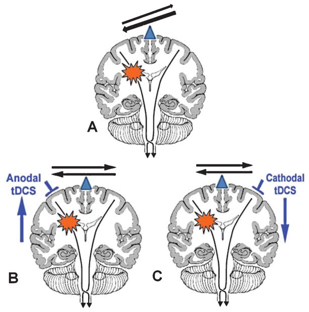
Purpose: It is thought that following a stroke the contralesional motor region exerts an undue inhibitory influence on the lesional motor region which might limit recovery. Pilot studies have shown that suppressing the contralesional motor region with cathodal transcranial Direct Current Stimulation (tDCS) can induce a short lasting functional benefit; greater and longer lasting effects might be achieved with combining tDCS with simultaneous occupational therapy (OT) and applying this intervention for multiple sessions.
Methods: We carried out a randomized, double blind, sham controlled study of chronic stroke patients receiving either 5 consecutive days of cathodal tDCS (for 30 minutes) applied to the contralesional motor region and simultaneous OT, or sham tDCS+OT.
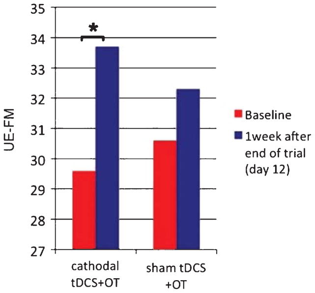
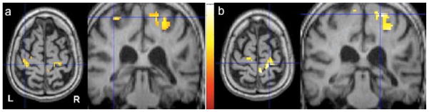
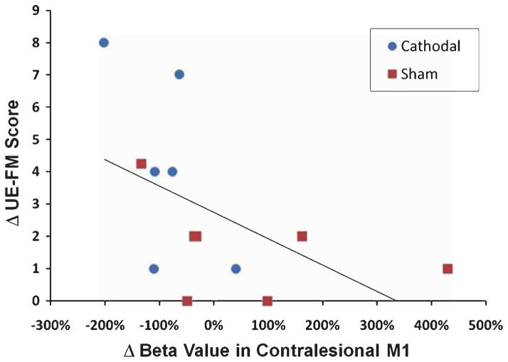
Results: we showed that cathodal tDCS+OT resulted in significantly more improvement in Range-Of-Motion in multiple joints of the paretic upper extremity and in the Upper-Extremity Fugl-Meyer scores than sham tDCS+OT, and that the effects lasted at least one week post-stimulation. Improvement in motor outcome scores was correlated with decrease in fMRI activation in the contralesional motor region exposed to cathodal stimulation.
Conclusions: This suggests that cathodal tDCS combined with OT leads to significant motor improvement after stroke due to a decrease in the inhibitory effect that the contralesional hemisphere exerts onto the lesional hemisphere.
Figures
References
-
- Adkins-Muir DL, Jones TA. Cortical electrical stimulation combined with rehabilitative training: Enhanced functional recovery and dendritic plasticity following focal cortical ischemia in rats. Neurol Res. 2003;25:780–788. - PubMed
-
- Baranyi A, Feher O. Synaptic facilitation requires paired activation of convergent pathways in the neocortex. Nature. 1981;290:413–415. - PubMed
-
- Boggio PS, Alonso-Alonso M, Mansur CG, Rigonatti SP, Schlaug G, et al. Hand function improvement with low-frequency repetitive transcranial magnetic stimulation of the unaffected hemisphere in a severe case of stroke. Am J Phys Med Rehabil. 2006;85:927–930. - PubMed
-
- Brett M, Leff AP, Rorden C, Ashburner J. Spatial normalization of brain images with focal lesions using cost function masking. NeuroImage. 2001;14:486–500. - PubMed
-
- Calautti C, Baron JC. Functional neuroimaging studies of motor recovery after stroke in adults: A review. Stroke. 2003;34:1553–1566. - PubMed
Publication types
MeSH terms
Substances
Grants and funding
LinkOut - more resources
Full Text Sources
Medical
