Controlled Clinical Trial
. 2012 Feb 15;590(4):919-36.
doi: 10.1113/jphysiol.2011.219592. Epub 2011 Nov 28.

The early release of planned movement by acoustic startle can be delayed by transcranial magnetic stimulation over the motor cortex

Affiliations
Controlled Clinical Trial

The early release of planned movement by acoustic startle can be delayed by transcranial magnetic stimulation over the motor cortex

Laila Alibiglou et al. J Physiol. .

Abstract

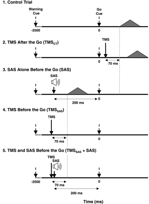
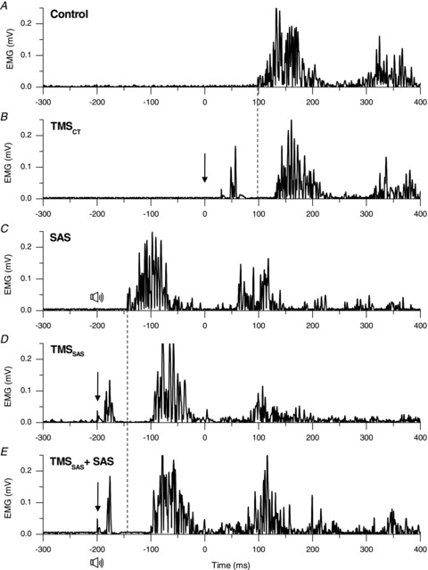
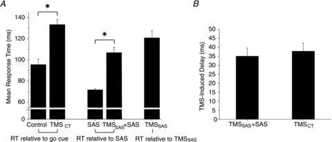
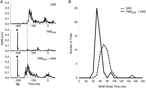
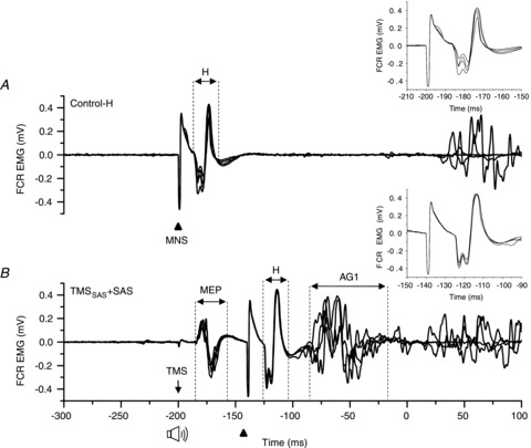
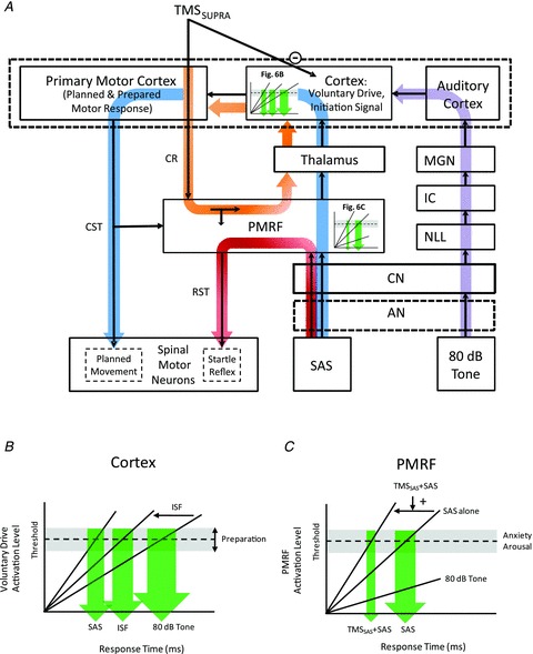
Previous studies have shown that preplanned movements can be rapidly released when a startling acoustic stimulus (SAS) is presented immediately prior to, or coincident with, the imperative signal to initiate movement. Based on the short latency of the onset of muscle activity (typically in less than 90 ms) and the frequent co-expression of startle responses in the neck and eye muscles, it has been proposed that the release of planned movements by a SAS is mediated by subcortical, possibly brainstem, pathways. However, a role for cortical structures in mediating these responses cannot be ruled out based on timing arguments alone. We examined the role of the cortex in the mediation of these responses by testing if a suprathreshold transcranial magnetic stimulation applied over the primary motor cortex, which suppresses voluntary drive and is known to delay movement initiation, could delay the release of movement by a SAS. Eight subjects performed an instructed-delay task requiring them to make a ballistic wrist movement to a target in response to an acoustic tone (control task condition). In a subset of trials subjects received one of the following: (1) suprathreshold TMS over the contralateral primary motor cortex 70 ms prior to their mean response time on control trials (TMS(CT)), (2) SAS 200 ms prior to the go cue (SAS), (3) suprathreshold TMS 70 ms prior to the mean SAS-evoked response time (TMS(SAS)), or (4) TMS(SAS) and SAS presented concurrently (TMS+SAS). Movement kinematics and EMG from the wrist extensors and flexors and sternocleidomastoid muscles were recorded. The application of TMS(CT) prior to control voluntary movements produced a significant delay in movement onset times (P < 0.001) (average delay = 37.7 ± 12.8 ms). The presentation of a SAS alone at -200 ms resulted in the release of the planned movement an average of 71.7 ± 2.7 ms after the startling stimulus. The early release of movement by a SAS was significantly delayed (P < 0.001, average delay = 35.0 ± 12.9 ms) when TMS(SAS) and SAS were presented concurrently. This delay could not be explained by a prolonged suppression of motor unit activity at the spinal level. These findings provide evidence that the release of targeted ballistic wrist movements by SAS is mediated, in part, by a fast conducting transcortical pathway via the primary motor cortex.


Figures

Figure 1
Figure 1. Diagram showing the timing of cues and stimuli for the five experimental conditions
Arrows indicate when TMS or SAS was applied during each condition. The filled triangles represent the EMG activity associated with the first agonist burst and the time that this burst was hypothesized to appear in each condition.
Figure 2
Figure 2. Representative trials across each experimental condition
Representative trials (AE) showing rectified EMG in the wrist extensor agonist muscle (extensor digitorum) in a single subject. The imperative go cue was presented at 0 ms. The downward arrow represents the timing of TMS and the speaker symbol represents the timing of the SAS. Note the delay in the onset of the agonist burst when TMS was applied immediately after the go cue (Fig. 2B) compared to the control trial (Fig. 2A). Similarly, note the delay in onset of the agonist burst (EMG onset =−103 ms) when TMS and SAS were applied together (Fig. 2E) at −200 ms compared to SAS alone at −200 ms (EMG onset =−140 ms).
Figure 3
Figure 3. Comparison of response times and TMS-induced delays
Mean and standard error for response time across the five experimental conditions (A) and the TMS-induced delay for the TMSCT and TMSSAS+SAS conditions (B). *Differences between conditions were significant (P < 0.05); RT, response time
Figure 4
Figure 4. Changes in the onset of SCM activity between the SAS alone and TMSSAS+ SAS conditions
A, three representative trials from a single subject showing rectified EMGs in the sternocleidomastoid (SCM) muscle for the SAS alone (left upper plot), the TMSSAS (left middle plot) and combined stimuli (TMSSAS+SAS) (left lower plot) conditions. Note that the latency of EMG activity was shortened when TMS was applied in combination with SAS. B, histogram of the number of trials observed with response times within 10 ms bins across five subjects. Note the shift in latency of the SCM burst and narrowing of the width of the distribution for the TMSSAS+SAS condition.
Figure 5
Figure 5. Comparison of the amplitude of the H-reflex between the Control-H and TMSSAS+SAS conditions
Example of EMG responses evoked in the flexor carpi radialis (FCR) muscle by electrical stimulation of the median nerve (MNS). Four consecutive trials in a single subject are shown in each plot. A, Control-H trials: the MNS was delivered at 200 ms before the go cue. B, TMSSAS+ SAS trials: the MNS was delivered 60 ms after TMS and SAS (during cortical silent period). Note that the latency and the amplitude of H-reflex (H) were similar in both conditions. The inset figures highlight the H-reflex size and shape across trials.
Figure 6
Figure 6. Model of the pathways mediating the release of movement in response to an 80 dB tone (Control trials), a SAS, TMSSAS alone and TMSSAS+SAS
This model shows two primary pathways (blue and purple) by which planned and prepared movements can be released by a sensory stimulus. The right portion of the model (purple line) shows the classic pathway by which a low intensity acoustic tone (e.g. 80 dB) travels to the auditory cortex. The auditory cortex then has input to a voluntary drive or initiating signal that triggers the release of the planned and prepared movement sequence at the level of the cortex. The timing and distribution of the response time is dependent upon the rate of rise of the input and the threshold to triggering the response, both of which can be modulated by intersensory facilitation or the level of preparation (Fig. 6B). Disruption of voluntary drive by suprathreshold TMS delays the release of the movement (TMSCT condition). The presentation of a SAS results in a synchronous afferent volley travelling to the PMRF and releasing a generalized startle reflex (SCM burst) via known pathways (shown in red). However, the timing of the generalized startle response can be altered by descending cortico-reticular input produced by TMS (orange line and Fig. 6C). The TMS-evoked volley to the PMRF increases the rise time of the startle triggering signal and produces both a reduction in startle onset latency and narrowing of the distribution of response times. Note that a decrease in threshold would not explain our results since this would not produce a narrowing of the response time distribution. Similar to the SAS response mechanism (shown in blue loop), the TMSSAS alone can trigger the early release of the planned movement through cortico-reticular inputs to the PMRF and a subsequent ascending volley via reticulo-thalamocortical pathways to the voluntary drive/initiation signal region of the cortex (shown in orange loop). Abbreviations: AN, auditory nerve; CN, cochlear nucleus; CR, corticoreticular projection; CST, corticospinal tract; IC, inferior colliculus; ISF, intersensory facilitation; MGN, medial geniculate nucleus; NLL, nuclei of the lateral lemniscus; PMRF, pontomedullary reticular formation; SAS, startling acoustic stimulus; SCM, sternocleidomastoid muscle; SUPRA, suprathreshold; RST, reticulospinal tract; TMS, transcranial magnetic stimulation.

References

    1. Buford JA, Davidson AG. Movement-related and preparatory activity in the reticulospinal system of the monkey. Exp Brain Res. 2004;159:284–300. - PubMed
    1. Carlsen AN, Chua R, Inglis JT, Sanderson DJ, Franks IM. Can prepared responses be stored subcortically? Exp Brain Res. 2004a;159:301–309. - PubMed
    1. Carlsen AN, Chua R, Inglis JT, Sanderson DJ, Franks IM. Prepared movements are elicited early by startle. J Mot Behav. 2004b;36:253–264. - PubMed
    1. Carlsen AN, Chua R, Inglis JT, Sanderson DJ, Franks IM. Differential effects of startle on reaction time for finger and arm movements. J Neurophysiol. 2009;101:306–314. - PMC - PubMed
    1. Carlsen AN, Dakin CJ, Chua R, Franks IM. Startle produces early response latencies that are distinct from stimulus intensity effects. Exp Brain Res. 2007;176:199–205. - PubMed

Publication types

LinkOut - more resources