

Action potential-triggered somatic exocytosis in mesencephalic trigeminal nucleus neurons in rat brain slices
- PMID: 22124145
- PMCID: PMC3381308
- DOI: 10.1113/jphysiol.2011.221051
Action potential-triggered somatic exocytosis in mesencephalic trigeminal nucleus neurons in rat brain slices
Abstract
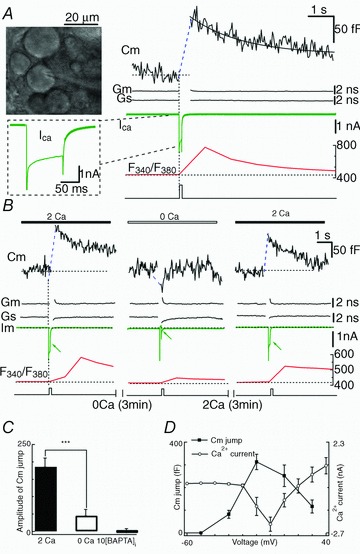
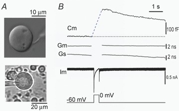
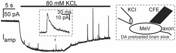
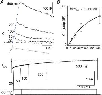
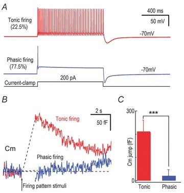
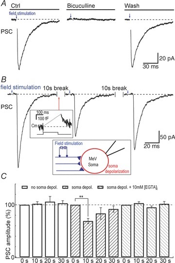
The neurons in the mesencephalic trigeminal nucleus (MeV) play essential roles in proprioceptive sensation of the face and oral cavity. The somata of MeV neurons are generally assumed to carry out neuronal functions but not to play a direct role in synaptic transmission. Using whole-cell recording and membrane capacitance (C(m)) measurements, we found that the somata of MeV neurons underwent robust exocytosis (C(m) jumps) upon depolarization and with the normal firing of action potentials in brain slices. Both removing [Ca(2+)](o) and buffering [Ca(2+)](i) with BAPTA blocked this exocytosis, indicating that it was completely Ca(2+) dependent. In addition, an electron microscopic study showed synaptic-like vesicles approximated to the plasma membrane in somata. There was a single Ca(2+)-dependent releasable vesicle pool with a peak release rate of 1912 fF s(-1). Importantly, following depolarization-induced somatic exocytosis, GABA-mediated postsynaptic currents were transiently reduced by 31%, suggesting that the somatic vesicular release had a retrograde effect on afferent GABAergic transmission. These results provide strong evidence that the somata of MeV neurons undergo robust somatic secretion and may play a crucial role in bidirectional communication between somata and their synaptic inputs in the central nervous system.
Figures
Similar articles
-
Ca(2+) and frequency dependence of exocytosis in isolated somata of magnocellular supraoptic neurones of the rat hypothalamus.J Physiol. 2004 Mar 16;555(Pt 3):699-711. doi: 10.1113/jphysiol.2003.051136. Epub 2003 Nov 28. J Physiol. 2004. PMID: 14645448 Free PMC article.
-
Excitatory GABAergic synaptic potentials in the mesencephalic trigeminal nucleus of adult rat in vitro.Neurosci Res. 2005 Apr;51(4):463-74. doi: 10.1016/j.neures.2004.12.016. Epub 2005 Jan 22. Neurosci Res. 2005. PMID: 15740809
-
Ca(2+)-dependent exocytosis in the somata of dorsal root ganglion neurons.Neuron. 1996 Jul;17(1):135-45. doi: 10.1016/s0896-6273(00)80287-1. Neuron. 1996. PMID: 8755485
-
L-type calcium channel-mediated plateau potentials in barrelette cells during structural plasticity.J Neurophysiol. 2002 Aug;88(2):794-801. doi: 10.1152/jn.2002.88.2.794. J Neurophysiol. 2002. PMID: 12163531 Free PMC article.
-
The Thermodynamically Expensive Contribution of Three Calcium Sources to Somatic Release of Serotonin.Int J Mol Sci. 2022 Jan 28;23(3):1495. doi: 10.3390/ijms23031495. Int J Mol Sci. 2022. PMID: 35163419 Free PMC article. Review.
Cited by
-
Extrasynaptic exocytosis and its mechanisms: a source of molecules mediating volume transmission in the nervous system.Front Physiol. 2012 Sep 4;3:319. doi: 10.3389/fphys.2012.00319. eCollection 2012. Front Physiol. 2012. PMID: 22969726 Free PMC article.
-
Search for unknown neural link between the masticatory and cognitive brain systems to clarify the involvement of its impairment in the pathogenesis of Alzheimer's disease.Front Cell Neurosci. 2024 Jun 27;18:1425645. doi: 10.3389/fncel.2024.1425645. eCollection 2024. Front Cell Neurosci. 2024. PMID: 38994328 Free PMC article. Review.
-
Brainstem neurons survive the identical ischemic stress that kills higher neurons: insight to the persistent vegetative state.PLoS One. 2014 May 6;9(5):e96585. doi: 10.1371/journal.pone.0096585. eCollection 2014. PLoS One. 2014. PMID: 24802253 Free PMC article.
-
Effect of amyloids on the vesicular machinery: implications for somatic neurotransmission.Philos Trans R Soc Lond B Biol Sci. 2015 Jul 5;370(1672):20140187. doi: 10.1098/rstb.2014.0187. Philos Trans R Soc Lond B Biol Sci. 2015. PMID: 26009766 Free PMC article. Review.
-
Veratridine modifies the gating of human voltage-gated sodium channel Nav1.7.Acta Pharmacol Sin. 2018 Nov;39(11):1716-1724. doi: 10.1038/s41401-018-0065-z. Epub 2018 Jun 27. Acta Pharmacol Sin. 2018. PMID: 29950616 Free PMC article.
References
Publication types
MeSH terms
Substances
LinkOut - more resources
Full Text Sources
Miscellaneous