. 2012 Feb;32(3):590-605.
doi: 10.1128/MCB.06570-11. Epub 2011 Nov 28.

Genetic reevaluation of the role of F-box proteins in cyclin D1 degradation

Affiliations

Genetic reevaluation of the role of F-box proteins in cyclin D1 degradation

Tomoharu Kanie et al. Mol Cell Biol. 2012 Feb.

Abstract

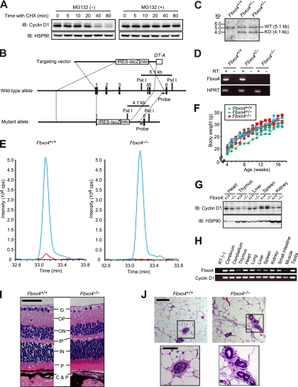
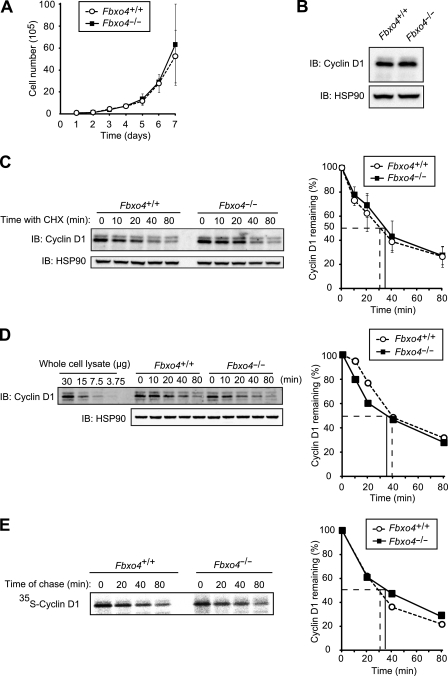
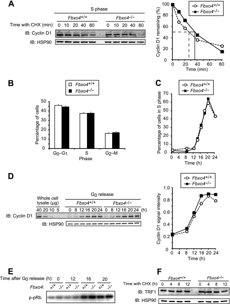
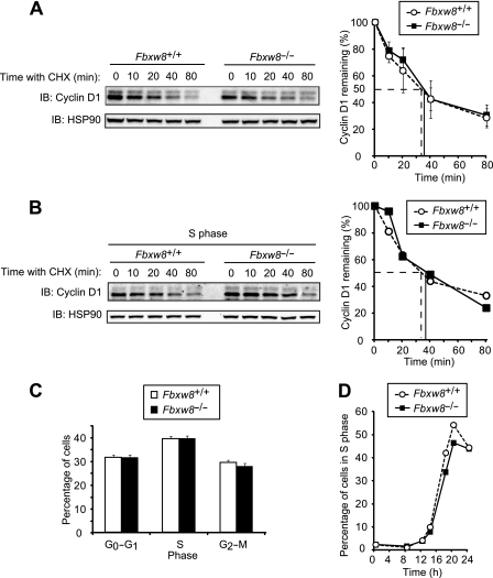
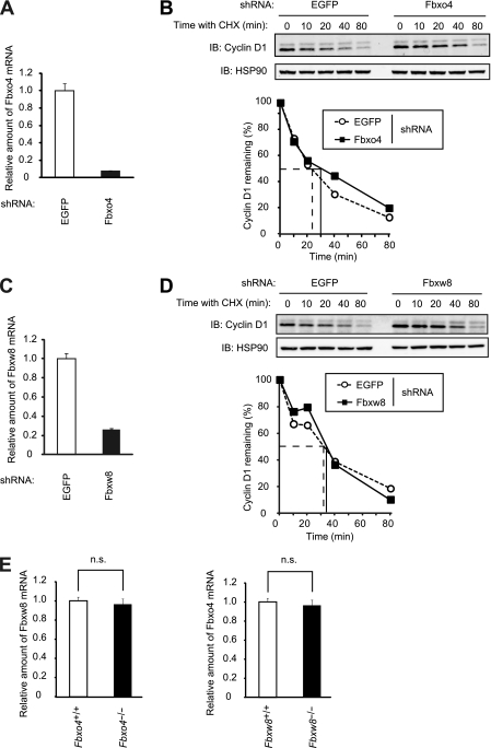
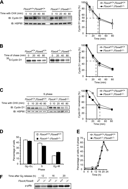
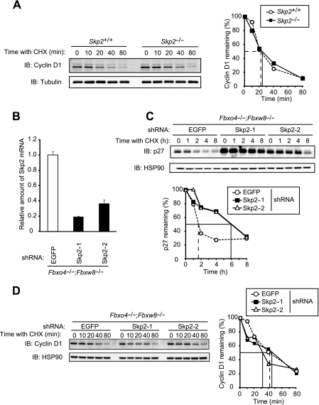
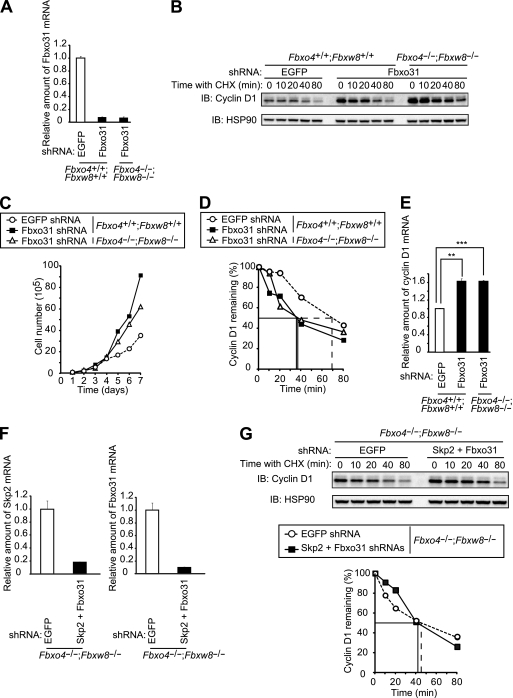
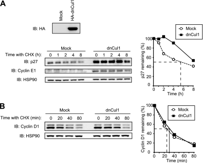
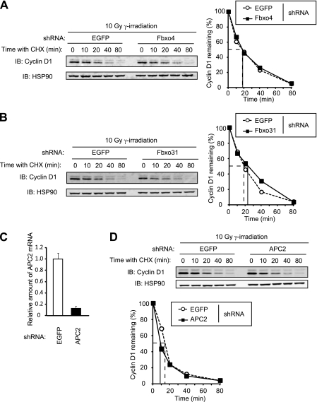
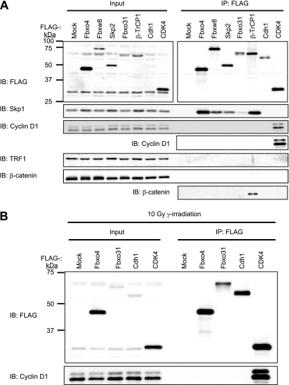
D-type cyclins play a pivotal role in G(1)-S progression of the cell cycle, and their expression is frequently deregulated in cancer. Cyclin D1 has a half-life of only ~30 min as a result of its ubiquitylation and proteasomal degradation, with various F-box proteins, including Fbxo4, Fbxw8, Skp2, and Fbxo31, having been found to contribute to its ubiquitylation. We have now generated Fbxo4-deficient mice and found no abnormalities in these animals. Cyclin D1 accumulation was thus not observed in Fbxo4(-/-) mouse tissues. The half-life of cyclin D1 in mouse embryonic fibroblasts (MEFs) prepared from Fbxo4(-/-), Fbxw8(-/-), and Fbxo4(-/-); Fbxw8(-/-) mice also did not differ from that in wild-type MEFs. Additional depletion of Skp2 and Fbxo31 in Fbxo4(-/-); Fbxw8(-/-) MEFs by RNA interference did not affect cyclin D1 stability. Although Fbxo31 depletion in MEFs increased cyclin D1 abundance, this effect appeared attributable to upregulation of cyclin D1 mRNA. Furthermore, abrogation of the function of the Skp1-Cul1-F-box protein (SCF) complex or the anaphase-promoting complex/cyclosome (APC/C) complexes did not alter the half-life of cyclin D1, whereas cyclin D1 degradation was dependent largely on proteasome activity. Our genetic analyses thus do not support a role for any of the four F-box proteins examined in cyclin D1 degradation during normal cell cycle progression. They suggest the existence of other ubiquitin ligases that target cyclin D1 for proteolysis.


Figures

FIG 1
FIG 1
Generation and phenotype of Fbxo4−/− mice. (A) Immunoblot (IB) analysis of cyclin D1 and HSP90 (loading control) in NIH 3T3 cells incubated with cycloheximide (CHX; 25 μg/ml) for the indicated times in the absence or presence of the proteasome inhibitor MG132 (10 μM). (B) Schematic representations of the wild-type mouse Fbxo4 locus, the targeting vector, and the mutant allele after homologous recombination. Exons are denoted by black boxes, and the positions of IRES-lacZ, PGK-neo-poly(A)-loxP (neo), and DT-A gene cassettes as well as PstI restriction sites are indicated. The expected sizes of DNA fragments that hybridize with the indicated probe in Southern blot analysis are shown. (C) Southern blot analysis of PstI-digested DNA from ES cells of the indicated Fbxo4 genotypes. The sizes of the hybridizing fragments corresponding to the wild-type (WT) and mutant (KO) alleles are indicated. (D) RT-PCR analysis of Fbxo4 and HPRT (control) mRNAs in Fbxo4+/+, Fbxo4+/−, and Fbxo4−/− MEFs. Reactions were performed with or without RT as indicated. (E) Extracted ion chromatograms for the Fbxo4 peptide in SRM analysis. The blue line indicates the signal for the internal standard (labeled with mTRAQ Δ4), and the red line indicates the signal for the endogenous Fbxo4 peptide (labeled with mTRAQ Δ0) for samples prepared from Fbxo4+/+ and Fbxo4−/− MEFs. (F) Representative growth curves for Fbxo4+/+, Fbxo4+/−, and Fbxo4−/− mice. (G) Immunoblot analysis of cyclin D1 and HSP90 in various tissues from 4-week-old Fbxo4+/+ or Fbxo4−/− mice. (H) RT-PCR analysis of Fbxo4 and cyclin D1 mRNAs in the indicated tissues of 12-week-old wild-type mice. (I) Histological analysis (hematoxylin-eosin staining) of the retina from 13-week-old Fbxo4−/− and wild-type littermates. G, ganglion cell layer; OP, outer plexiform layer; ON, outer nuclear layer; IP, inner plexiform layer; IN, inner nuclear layer; P, photoreceptor cell layer; C&P, choroid and pigment cells. Scale bar, 50 μm. (J) Histological analysis (hematoxylin-eosin staining) of the mammary gland from 20-week-old nulliparous Fbxo4−/− and wild-type littermates. The boxed areas in the upper panels are shown at a higher magnification in the lower panels; scale bars, 100 and 50 μm, respectively.
FIG 2
FIG 2
Cyclin D1 does not accumulate in Fbxo4−/− MEFs. (A) Growth curves for Fbxo4+/+ and Fbxo4−/− MEFs. Data are means ± SD of values obtained from three MEF preparations of each genotype. (B) Immunoblot analysis of cyclin D1 in MEFs derived from Fbxo4+/+ and Fbxo4−/− mice. (C) Immunoblot analysis of cyclin D1 (left) and its quantification (right) for Fbxo4+/+ and Fbxo4−/− MEFs incubated with cycloheximide (25 μg/ml) for the indicated times. The half-life of cyclin D1 in cells of each genotype is indicated. Quantitative data are means ± SD of values obtained from four MEF preparations of each genotype. (D) Quantitative immunoblot analysis of cyclin D1 with fluorescent secondary antibodies (left) and its quantification (right) for Fbxo4+/+ and Fbxo4−/− MEFs incubated with cycloheximide for the indicated times. The indicated amounts of whole-cell lysate were analyzed as a standard. (E) Pulse-chase analysis of cyclin D1 (left) and its quantification (right) for Fbxo4+/+ and Fbxo4−/− MEFs. Cells were metabolically labeled with [35S]methionine and [35S]cysteine for 1 h and then chased with nonradioactive methionine and cysteine for the indicated times. Cell lysates were then prepared and subjected to immunoprecipitation with antibodies to cyclin D1, and the resulting precipitates were subjected to SDS-PAGE and autoradiography.
FIG 3
FIG 3
Depletion of Fbxo4 does not affect cyclin D1 function. (A) Immunoblot analysis of cyclin D1 (left) and its quantification (right) for Fbxo4+/+ and Fbxo4−/− MEFs incubated with cycloheximide in S phase. Cells were released from G0 phase and cultured for 20 h (at which time most cells were in S phase) before incubation with cycloheximide (25 μg/ml) for the indicated times. (B) Cell cycle distribution of asynchronous Fbxo4+/+ and Fbxo4−/− MEFs as determined by flow cytometry. Data are means ± SD of results from three independent experiments. (C) Kinetics of the reentry of Fbxo4+/+ and Fbxo4−/− MEFs into the cell cycle from G0 phase. Data are representative of three independent experiments. (D) Fluorescence-based quantitative immunoblot analysis of cyclin D1 (left) and its quantification (right) for Fbxo4+/+ and Fbxo4−/− MEFs released from G0 phase for the indicated times. The indicated amounts of whole-cell lysate were analyzed as a standard. A signal intensity of 1.0 corresponds to that for 40 μg of cell lysate. (E) In vitro CDK4 kinase assay for Fbxo4+/+ and Fbxo4−/− MEFs released from G0 phase for the indicated times. Cell lysates were subjected to immunoprecipitation with antibodies to CDK4, and the resulting precipitates were assayed for CDK4 kinase activity with a glutathione S-transferase (GST)-pRb fusion protein as substrate. (F) Immunoblot analysis of TRF1 in Fbxo4+/+ and Fbxo4−/− MEFs incubated with cycloheximide for the indicated times.
FIG 4
FIG 4
Cyclin D1 does not accumulate in Fbxw8−/− MEFs. (A) Immunoblot analysis of cyclin D1 (left) and its quantification (right) for MEFs from Fbxw8+/+ and Fbxw8−/− mice incubated with cycloheximide (25 μg/ml) for the indicated times. Quantitative data are means ± SD of values obtained from four MEF preparations of each genotype. (B) Immunoblot analysis of cyclin D1 (left) and its quantification (right) for Fbxw8+/+ and Fbxw8−/− MEFs incubated with cycloheximide for the indicated times in S phase. (C) Cell cycle distribution of asynchronous Fbxw8+/+ and Fbxw8−/− MEFs. Data are means ± SD of results from four independent experiments. (D) Kinetics of the reentry of Fbxw8+/+ and Fbxw8−/− MEFs into the cell cycle from G0 phase. Data are representative of results from three independent experiments.
FIG 5
FIG 5
Acute depletion of Fbxo4 or Fbxw8 does not affect the half-life of cyclin D1. (A) Quantitative RT-PCR analysis of Fbxo4 mRNA in NIH 3T3 cells infected with a retroviral vector encoding control (EGFP) or Fbxo4 shRNAs. Data are means ± SD of triplicates from a representative experiment. (B) Immunoblot analysis of cyclin D1 (top) and its quantification (bottom) for NIH 3T3 cells infected as described for panel A and incubated with cycloheximide (25 μg/ml) for the indicated times. (C) Quantitative RT-PCR analysis of Fbxw8 mRNA in NIH 3T3 cells infected with a retroviral vector encoding control (EGFP) or Fbxw8 shRNAs. Data are means ± SD of triplicates from a representative experiment. (D) Immunoblot analysis of cyclin D1 (top) and its quantification (bottom) for NIH 3T3 cells infected as described for panel C and incubated with cycloheximide for the indicated times. (E) Quantitative RT-PCR analysis of Fbxw8 mRNA in Fbxo4+/+ and Fbxo4−/− MEFs and of Fbxo4 mRNA in Fbxw8+/+ and Fbxw8−/− MEFs. Data are means ± SD of triplicates from a representative experiment. n.s., not significant (Student's unpaired t test).
FIG 6
FIG 6
Cyclin D1 does not accumulate in Fbxo4−/−; Fbxw8−/− MEFs. (A) Immunoblot analysis of cyclin D1 (left) and its quantification (right) for Fbxo4+/+; Fbxw8+/+ and Fbxo4−/−; Fbxw8−/− MEFs incubated with cycloheximide (25 μg/ml) for the indicated times. Quantitative data are means ± SD of values obtained from five MEF preparations of each genotype. (B) Pulse-chase analysis of cyclin D1 (left) and its quantification (right) for Fbxo4+/+; Fbxw8+/+ and Fbxo4−/−; Fbxw8−/− MEFs. (C) Immunoblot analysis of cyclin D1 (left) and its quantification (right) for Fbxo4+/+; Fbxw8+/+ and Fbxo4−/−; Fbxw8−/− MEFs incubated with cycloheximide for the indicated times in S phase. (D) Cell cycle distribution of asynchronous Fbxo4+/+; Fbxw8+/+ and Fbxo4−/−; Fbxw8−/− MEFs. Data are means ± SD from four independent experiments. (E) Kinetics of the reentry of Fbxo4+/+; Fbxw8+/+ and Fbxo4−/−; Fbxw8−/− MEFs into the cell cycle from G0 phase. Data are representative of three independent experiments. (F) In vitro CDK4 kinase assay for Fbxo4+/+; Fbxw8+/+ and Fbxo4−/−; Fbxw8−/− MEFs released from G0 phase for the indicated times.
FIG 7
FIG 7
Cyclin D1 does not accumulate in Skp2-depleted Fbxo4−/−; Fbxw8−/− MEFs. (A) Immunoblot analysis of cyclin D1 (left) and its quantification (right) for MEFs from Skp2+/+ and Skp2−/− mice incubated with cycloheximide (25 μg/ml) for the indicated times. α-Tubulin was examined as a loading control. (B) Quantitative RT-PCR analysis of Skp2 mRNA in Fbxo4−/−; Fbxw8−/− MEFs infected with a retroviral vector encoding control (EGFP) or one of two Skp2 shRNAs. Data are means ± SD of triplicates from a representative experiment. (C) Immunoblot analysis of p27 (top) and its quantification (bottom) for MEFs infected as described for panel B and incubated with cycloheximide for the indicated times. (D) Immunoblot analysis of cyclin D1 (left) and its quantification (right) for MEFs infected as described for panel B and incubated with cycloheximide for the indicated times.
FIG 8
FIG 8
Cyclin D1 is degraded in the absence of Fbxo4, Fbxw8, Skp2, and Fbxo31. (A) Quantitative RT-PCR analysis of Fbxo31 mRNA in Fbxo4+/+; Fbxw8+/+ and Fbxo4−/−; Fbxw8−/− MEFs infected with a retroviral vector encoding EGFP or Fbxo31 shRNAs. Data are means ± SD of triplicates from a representative experiment. (B) Immunoblot analysis of cyclin D1 in MEFs as described for panel A incubated with cycloheximide (25 μg/ml) for the indicated times. (C) Growth curves for MEFs as described for panel A. Data are representative of results from three independent experiments. (D) Quantification of cyclin D1 abundance in the experiment shown in panel B. (E) Quantitative RT-PCR analysis of cyclin D1 mRNA in MEFs as described for panel A. Data are means ± SD of triplicates from a representative experiment. **, P < 0.005; ***, P < 0.0005 (Student's unpaired t test). (F) Quantitative RT-PCR analysis of Skp2 and Fbxo31 mRNAs in Fbxo4−/−; Fbxw8−/− MEFs infected with retroviral vectors encoding EGFP or both Skp2 and Fbxo31 shRNAs. Data are means ± SD of triplicates from a representative experiment. (G) Immunoblot analysis of cyclin D1 (top) and its quantification (bottom) for MEFs, as described for panel F, incubated with cycloheximide for the indicated times.
FIG 9
FIG 9
Inhibition of Cul1 function does not affect the stability of cyclin D1. (A) Immunoblot analysis with antibodies to the hemagglutinin epitope (HA; top) or to p27 and cyclin E1 (bottom left), as well as quantification of p27 (bottom right), for NIH 3T3 cells infected with a retroviral vector for an HA-tagged dominant negative mutant (dn) of Cul1 (or subjected to mock infection) and exposed to cycloheximide (25 μg/ml) for the indicated times. (B) Immunoblot analysis of cyclin D1 (left) and its quantification (right) for cells as described for panel A exposed to cycloheximide for the indicated times.
FIG 10
FIG 10
The absence of Fbxo4, Fbxo31, or APC2 does not affect the stability of cyclin D1 after DNA damage. (A) Immunoblot analysis of cyclin D1 (left) and its quantification (right) for NIH 3T3 cells infected with a retroviral vector encoding control (EGFP) or Fbxo4 shRNAs. Cells were incubated with cycloheximide (25 μg/ml) for the indicated times beginning 1 h after gamma irradiation (10 Gy). (B) Immunoblot analysis of cyclin D1 (left) and its quantification (right) for NIH 3T3 cells infected with a retroviral vector encoding EGFP or Fbxo31 shRNAs and treated as described for panel A. (C) Quantitative RT-PCR analysis of APC2 mRNA in NIH 3T3 cells infected with a retroviral vector encoding EGFP or APC2 shRNAs. Data are means ± SD of triplicates from a representative experiment. (D) Immunoblot analysis of cyclin D1 (top) and its quantification (bottom), as described for panel C, for cells incubated with cycloheximide for the indicated times beginning 1 h after gamma irradiation.
FIG 11
FIG 11
Interactions between ubiquitin ligases and cyclin D1. (A) Lysates of NIH 3T3 cells infected with a retroviral vector encoding FLAG-tagged Fbxo4, Fbxw8, Skp2, Fbxo31, β-TrCP1, Cdh1, or CDK4 or subjected to mock infection were subjected to immunoprecipitation (IP) with antibodies to FLAG, and the resulting precipitates as well as the original lysates (Input) were subjected to immunoblot analysis with antibodies to FLAG or to the indicated proteins. (B) NIH 3T3 cells infected with a retroviral vector encoding FLAG-tagged Fbxo4, Fbxo31, Cdh1, or CDK4 were subjected to gamma irradiation (10 Gy). One hour after irradiation, the cells were lysed and subjected to immunoprecipitation with antibodies to FLAG, and the resulting precipitates as well as the original lysates were subjected to immunoblot analysis with antibodies to FLAG or to cyclin D1.

References

    1. Agami R, Bernards R. 2000. Distinct initiation and maintenance mechanisms cooperate to induce G1 cell cycle arrest in response to DNA damage. Cell 102:55–66 - PubMed
    1. Alt JR, Cleveland JL, Hannink M, Diehl JA. 2000. Phosphorylation-dependent regulation of cyclin D1 nuclear export and cyclin D1-dependent cellular transformation. Genes Dev. 14:3102–3114 - PMC - PubMed
    1. Ang XL, Wade Harper J. 2005. SCF-mediated protein degradation and cell cycle control. Oncogene 24:2860–2870 - PubMed
    1. Baldin V, Lukas J, Marcote MJ, Pagano M, Draetta G. 1993. Cyclin D1 is a nuclear protein required for cell cycle progression in G1. Genes Dev. 7:812–821 - PubMed
    1. Barbash O, et al. 2008. Mutations in Fbx4 inhibit dimerization of the SCFFbx4 ligase and contribute to cyclin D1 overexpression in human cancer. Cancer Cell 14:68–78 - PMC - PubMed

Publication types

LinkOut - more resources