. 2012 Jan;30(1):87-96.
doi: 10.1097/HJH.0b013e32834dde5f.

Telmisartan ameliorates lipopolysaccharide-induced innate immune response through peroxisome proliferator-activated receptor-γ activation in human monocytes

Affiliations

Telmisartan ameliorates lipopolysaccharide-induced innate immune response through peroxisome proliferator-activated receptor-γ activation in human monocytes

Tao Pang et al. J Hypertens. 2012 Jan.

Abstract

Objective: Angiotensin II type 1 receptor (AT1) blockers (ARBs) reduce the bacterial endotoxin lipopolysaccharide (LPS)-induced innate immune response in human circulating monocytes expressing few AT1. To clarify the mechanisms of anti-inflammatory effects of ARBs with different peroxisome proliferator-activated receptor-γ (PPARγ)-activating potencies, we focused our study on telmisartan, an ARB with the highest PPARγ-stimulating activity.

Methods: Human circulating monocytes and monocytic THP-1 (human acute monocytic leukemia cell line) cells were exposed to 50 ng/ml LPS with or without pre-incubation with telmisartan. AT1 mRNA and protein expressions were determined by real-time PCR and membrane receptor binding assay, respectively. The expression of pro-inflammatory factors was determined by real-time PCR, western blot analysis and ELISA. PPARγ activation was measured by electrophoretic mobility shift assay and its role was determined by pharmacological inhibition and PPARγ gene silencing.

Results: In human monocytes, telmisartan significantly attenuated the LPS-induced expression of pro-inflammatory factors, the release of pro-inflammatory cytokines and prostaglandin E2, nuclear factor-κB activation and reactive oxygen species formation. In THP-1 cells, telmisartan significantly reduced LPS-induced tumor necrosis factor-α, inhibitor of κB-α, monocyte chemotactic protein-1 (MCP-1) and lectin-like oxidized low-density lipoprotein receptor-1 gene expression and MCP-1-directed migration. Telmisartan also stimulated the expression of the PPARγ target genes cluster of differentiation 36 and ATP-binding cassette subfamily G member 1 in monocytes. The anti-inflammatory effects of telmisartan were prevented by both PPARγ antagonism and PPARγ gene silencing. Anti-inflammatory effects of ARBs correlated with their PPARγ agonist potency.

Conclusion: Our observations demonstrate that in human monocytes, ARBs inhibit the LPS-induced pro-inflammatory response to a major extent through the PPARγ activation pathway and may be beneficial for the treatment of cardiovascular and metabolic disorders in which inflammation plays a major role.


Conflict of interest statement

Conflicts of interest

There are no conflicts of interest.

Figures

Fig. 1
Fig. 1
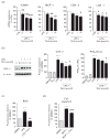
Telmisartan (Telm) inhibited lipopolysaccharide (LPS)-induced pro-inflammatory cytokine release and mRNA expression in human monocytes. Monocytes were incubated with 50 ng/ml LPS alone or after 2 h pre-incubation with different doses of Telm. (a) Cumulative release of pro-inflammatory cytokines 4 h after LPS addition as determined by ELISA. (b) Expression of cytokine mRNA after 2 h incubation with LPS as determined by real-time PCR. Results are means ± SEM from three independent experiments and are expressed as a percentage of LPS-induced response. IL, interleukin; TNF, tumor necrosis factor. #P<0.05; ##P<0.01; ###P<0.001 compared with LPS.
Fig. 2
Fig. 2
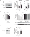
Telmisartan (Telm) inhibited lipopolysaccharide (LPS)-induced pro-inflammatory responses in human monocytes. Human monocytes were pre-incubated for 2 h with vehicle (DMSO) or Telm and subsequently exposed to 50 ng/ml LPS. (a) Expression of pro-inflammatory marker mRNA 2 h after LPS addition. (b) Cyclooxygenase-2 (COX-2) protein expression and cumulative prostaglandin E2 (PGE2) release in monocytes exposed to LPS for 24 h as determined by western blot and EIA, respectively. COX-2 protein expression was determined by western blot and quantified by densitometry. Values are presented as percentage of DMSO-treated group, as means ± SEM from at least three independent experiments. ***P<0.001 vs. DMSO; #P<0.05 vs. LPS. Picture shows a representative western blot. (c) Reactive oxygen species (ROS) production was determined by dichlorodihydrofluorescein diacetate probe oxidation in monocytes pretreated with vehicle or 10 μmol/l Telm and then exposed to LPS for 2 h. (d) Cell migration assay was performed on THP-1 monocytic cells pretreated with vehicle or 10 μmol/l Telm and subsequently stimulated with 100 ng/ml monocyte chemotactic protein-1 (MCP-1) for 4 h. Results are means ± SEM from at least three independent experiments. **P<0.01 vs. DMSO; ***P<0.001 vs. DMSO; #P<0.05 vs. LPS; ##P<0.01 vs. LPS; ###P<0.001 vs. LPS or MCP-1. ICAM-1, intercellular adhesion molecule 1; LOX-1, lectin-like oxidized low-density lipoprotein receptor-1.
Fig. 3
Fig. 3
Anti-inflammatory effects of telmisartan (Telm) in human monocytes involved inhibition of mitogen-activated protein kinase (MAPK) and nuclear factor-κB (NF-κB) activation. Human monocytes were pre-incubated for 2 h with vehicle (DMSO) or 10 μmol/l Telm and subsequently exposed to lipopolysaccharide (LPS) (50 ng/ml). (a) Representative western blot image showing inhibitory effect of Telm on LPS-induced phosphorylation of p38 MAPK and extracellular signal-regulated kinases 1/2 (ERK1/2) in monocytes incubated for 30 min with LPS. Quantitative densitometric values are presented as percentage of DMSO-treated group, and as means ± SEM from at least three independent experiments. **P<0.01 vs. DMSO; ***P<0.001 vs. DMSO; ##P<0.01 vs. LPS; ###P<0.001 vs. LPS. (b) Telm dose dependently inhibited inhibitor of κB-α (IκB-α) mRNA expression induced by 2 h incubation with LPS, and prevented LPS-induced IκB-α protein degradation time dependently. IκB-α protein levels are presented as means ± SEM from three independent experiments. *P<0.05 vs. control (0 h); #P<0.05 vs. LPS with Telm group. Pictures are representative western blots. (c) Nuclear proteins extracted from human monocytes treated with LPS with or without Telm were used to measure NF-κB p65 protein expression by western blot. Representative western blot is shown. NF-κB p65 protein values are presented as means ± SEM from three independent experiments. ***P<0.001 vs. DMSO; ###P<0.001 vs. LPS.
Fig. 4
Fig. 4
Telmisartan (Telm) activated peroxisome proliferator-activated receptor-γ (PPARγ) and increased PPARγ target gene expression in human monocytes and THP-1 cells. Primary human monocytes or THP-1 cells were incubated for 4 h with vehicle (DMSO) or 10 μmol/l Telm. PPARγ activation was analyzed by electrophoretic mobility shift assay. Representative pictures show PPARγ–DNA binding in human monocytes (a) and THP-1 cells (b), and the intensity of each band was measured by densitometry for quantitative analysis. Anti-PPARγ antibody (2 μg) and a 100-fold excess of cold probe were used to determine the specificity of the shift. Pre-incubation of nuclear extracts from Telm-treated cells with an anti-PPARγ antibody led to a profound decrease in PPARγ–DNA binding activity, demonstrating the reaction specificity. Expression of mRNA of PPARγ and PPARγ target genes cluster of differentiation 36 (CD36) and ATP-binding cassette subfamily G member 1 (ABCG1) in monocytes (c) and THP-1 cells (d) was determined by real-time PCR. Results are means ± SEM of three independent experiments. *P<0.05 vs. DMSO; ***P<0.001 vs. DMSO.
Fig. 5
Fig. 5
Peroxisome proliferator-activated receptor-γ (PPARγ) gene silencing eliminated the effect of telmisartan (Telm) on lipopolysaccharide (LPS)-induced gene expression in THP-1 cells. Human PPARγ-specific or nontargeting scrambled control small interfering RNA (siRNA) were transfected into THP-1 cells. PPARγ protein levels were measured at 48 and 72 h posttransfection by western blot (a). PPARγ mRNA expression (b) and cluster of differentiation 36 (CD36) and ATP-binding cassette subfamily G member 1 (ABCG1) mRNA expression (c) were measured at 72 h of posttransfection by real-time PCR. ***P<0.001 vs. scrambled siRNA or DMSO; ###P<0.001 vs. Telm. The inflammatory markers tumor necrosis factor-α (TNFα) and inhibitor of κB-α (IκB-α) were measured in cells treated for 2 h with LPS (50 ng/ml) alone or in a combination with 10 μmol/l Telm with and without PPARγ siRNA transfection (d). Results are means ± SEM from three independent experiments. ###P<0.001 vs. LPS; ***P<0.001 vs. LPS with Telm.
Fig. 6
Fig. 6
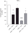
Peroxisome proliferator-activated receptor-γ (PPARγ) antagonist prevents telmisartan (Telm) amelioration of lipopolysaccharide (LPS)-induced tumor necrosis factor-α (TNFα) release. Monocytes pretreated for 1 h with 1 μmol/l PPARγ antagonist T0070907 (T007) were incubated for 1 h with 10 μmol/l Telm or 10 μmol/l PPARγ agonist troglitazone (Tgz) followed by 4 h incubation in the presence of 50 ng/ml LPS. Cumulative TNFα release was determined in culture medium by ELISA. Results are means ± SEM of three independent experiments and are expressed as a percentage of LPS-induced response. #P<0.05 vs. LPS; ###P<0.001 vs. LPS; *P<0.05 vs. LPS with Telm or LPS with Tgz.

Similar articles

Cited by

References

    1. Timmermans PB, Wong PC, Chiu AT, Herblin WF, Benfield P, Carini DJ, et al. Angiotensin II receptors and angiotensin II receptor antagonists. Pharmacol Rev. 1993;45:205–251. - PubMed
    1. Chrysant SG, Chrysant GS, Chrysant C, Shiraz M. The treatment of cardiovascular disease continuum: focus on prevention and RAS blockade. Curr Clin Pharmacol. 2010;5:89–95. - PubMed
    1. Bakris G. Are there effects of renin-angiotensin system antagonists beyond blood pressure control? Am J Cardiol. 2010;105:21A–29A. - PubMed
    1. Konstam MA, Neaton JD, Dickstein K, Drexler H, Komajda M, Martinez FA, et al. Effects of high-dose versus low-dose losartan on clinical outcomes in patients with heart failure (HEAAL study): a randomised, double-blind trial. Lancet. 2009;374:1840–1848. - PubMed
    1. Neldam S. Choosing an angiotensin-receptor blocker: blood pressure lowering, cardiovascular protection or both? Future Cardiol. 2010;6:129–135. - PubMed

Publication types