Tail-anchor targeting by a Get3 tetramer: the structure of an archaeal homologue
- PMID: 22124326
- PMCID: PMC3273380
- DOI: 10.1038/emboj.2011.433
Tail-anchor targeting by a Get3 tetramer: the structure of an archaeal homologue
Abstract
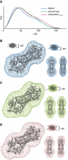
Efficient delivery of membrane proteins is a critical cellular process. The recently elucidated GET (Guided Entry of TA proteins) pathway is responsible for the targeted delivery of tail-anchored (TA) membrane proteins to the endoplasmic reticulum. The central player is the ATPase Get3, which in its free form exists as a dimer. Biochemical evidence suggests a role for a tetramer of Get3. Here, we present the first crystal structure of an archaeal Get3 homologue that exists as a tetramer and is capable of TA protein binding. The tetramer generates a hydrophobic chamber that we propose binds the TA protein. We use small-angle X-ray scattering to provide the first structural information of a fungal Get3/TA protein complex showing that the overall molecular envelope is consistent with the archaeal tetramer structure. Moreover, we show that this fungal tetramer complex is capable of TA insertion. This allows us to suggest a model where a tetramer of Get3 sequesters a TA protein during targeting to the membrane.
Conflict of interest statement
The authors declare that they have no conflict of interest.
Figures


Similar articles
-
Get1 stabilizes an open dimer conformation of get3 ATPase by binding two distinct interfaces.J Mol Biol. 2012 Sep 21;422(3):366-75. doi: 10.1016/j.jmb.2012.05.045. Epub 2012 Jun 7. J Mol Biol. 2012. PMID: 22684149
-
Protein targeting. Structure of the Get3 targeting factor in complex with its membrane protein cargo.Science. 2015 Mar 6;347(6226):1152-5. doi: 10.1126/science.1261671. Science. 2015. PMID: 25745174 Free PMC article.
-
The crystal structures of yeast Get3 suggest a mechanism for tail-anchored protein membrane insertion.PLoS One. 2009 Nov 30;4(11):e8061. doi: 10.1371/journal.pone.0008061. PLoS One. 2009. PMID: 19956640 Free PMC article.
-
Structures of Get3, Get4, and Get5 provide new models for TA membrane protein targeting.Structure. 2010 Aug 11;18(8):897-902. doi: 10.1016/j.str.2010.07.003. Structure. 2010. PMID: 20696390 Free PMC article. Review.
-
Endoplasmic reticulum targeting and insertion of tail-anchored membrane proteins by the GET pathway.Cold Spring Harb Perspect Biol. 2013 Aug 1;5(8):a013334. doi: 10.1101/cshperspect.a013334. Cold Spring Harb Perspect Biol. 2013. PMID: 23906715 Free PMC article. Review.
Cited by
-
In vitro Assays for Targeting and Insertion of Tail-Anchored Proteins Into the ER Membrane.Curr Protoc Cell Biol. 2018 Dec;81(1):e63. doi: 10.1002/cpcb.63. Epub 2018 Sep 25. Curr Protoc Cell Biol. 2018. PMID: 30253068 Free PMC article.
-
Differential gradients of interaction affinities drive efficient targeting and recycling in the GET pathway.Proc Natl Acad Sci U S A. 2014 Nov 18;111(46):E4929-35. doi: 10.1073/pnas.1411284111. Epub 2014 Nov 3. Proc Natl Acad Sci U S A. 2014. PMID: 25368153 Free PMC article.
-
Loss of GET pathway orthologs in Arabidopsis thaliana causes root hair growth defects and affects SNARE abundance.Proc Natl Acad Sci U S A. 2017 Feb 21;114(8):E1544-E1553. doi: 10.1073/pnas.1619525114. Epub 2017 Jan 17. Proc Natl Acad Sci U S A. 2017. PMID: 28096354 Free PMC article.
-
Get3 is a holdase chaperone and moves to deposition sites for aggregated proteins when membrane targeting is blocked.J Cell Sci. 2013 Jan 15;126(Pt 2):473-83. doi: 10.1242/jcs.112151. Epub 2012 Nov 30. J Cell Sci. 2013. PMID: 23203805 Free PMC article.
-
A portrait of the GET pathway as a surprisingly complicated young man.Trends Biochem Sci. 2012 Oct;37(10):411-7. doi: 10.1016/j.tibs.2012.07.004. Epub 2012 Aug 30. Trends Biochem Sci. 2012. PMID: 22951232 Free PMC article. Review.
References
-
- Adams PD, Grosse-Kunstleve RW, Hung LW, Ioerger TR, McCoy AJ, Moriarty NW, Read RJ, Sacchettini JC, Sauter NK, Terwilliger TC (2002) PHENIX: building new software for automated crystallographic structure determination. Acta Cryst D 58: 1948–1954 - PubMed
-
- Beilharz T, Egan B, Silver PA, Hofmann K, Lithgow T (2003) Bipartite signals mediate subcellular targeting of tail-anchored membrane proteins in Saccharomyces cerevisiae. J Biol Chem 278: 8219–8223 - PubMed
-
- Bernstein HD, Poritz MA, Strub K, Hoben PJ, Brenner S, Walter P (1989) Model for signal sequence recognition from amino-acid sequence of 54K subunit of signal recognition particle. Nature 340: 482–486 - PubMed
Publication types
MeSH terms
Substances
Associated data
- Actions
- Actions
Grants and funding
LinkOut - more resources
Full Text Sources
Molecular Biology Databases

