. 2012 Feb 1;31(3):754-66.
doi: 10.1038/emboj.2011.429. Epub 2011 Nov 29.

ACBD3-mediated recruitment of PI4KB to picornavirus RNA replication sites

Affiliations

ACBD3-mediated recruitment of PI4KB to picornavirus RNA replication sites

Jun Sasaki et al. EMBO J. .

Abstract

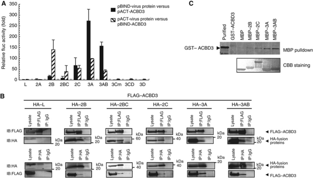
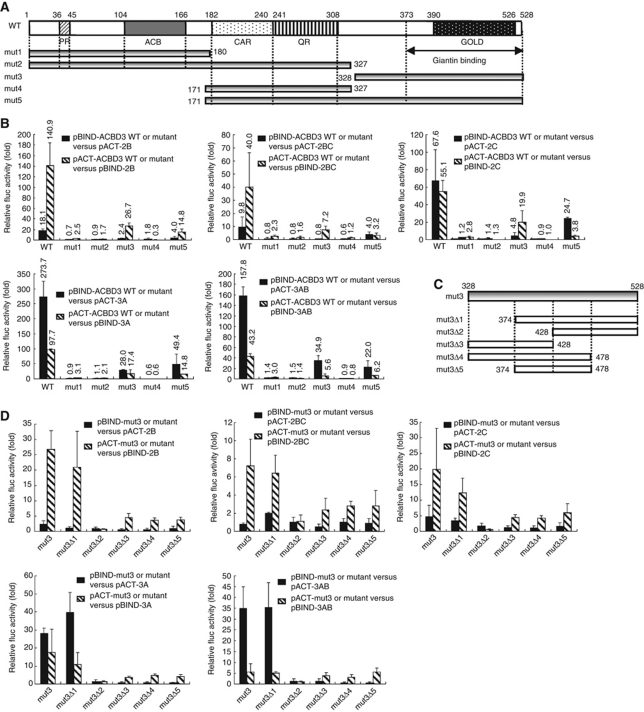
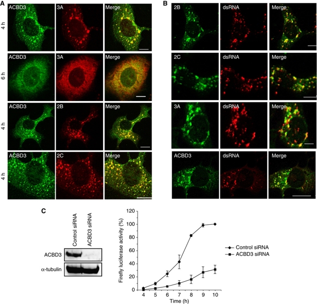
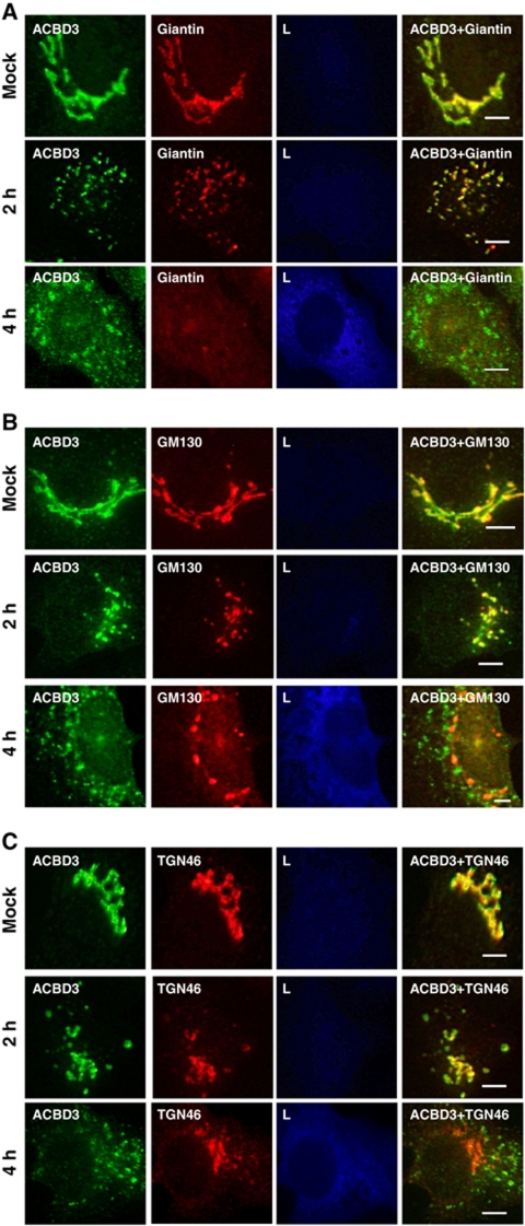
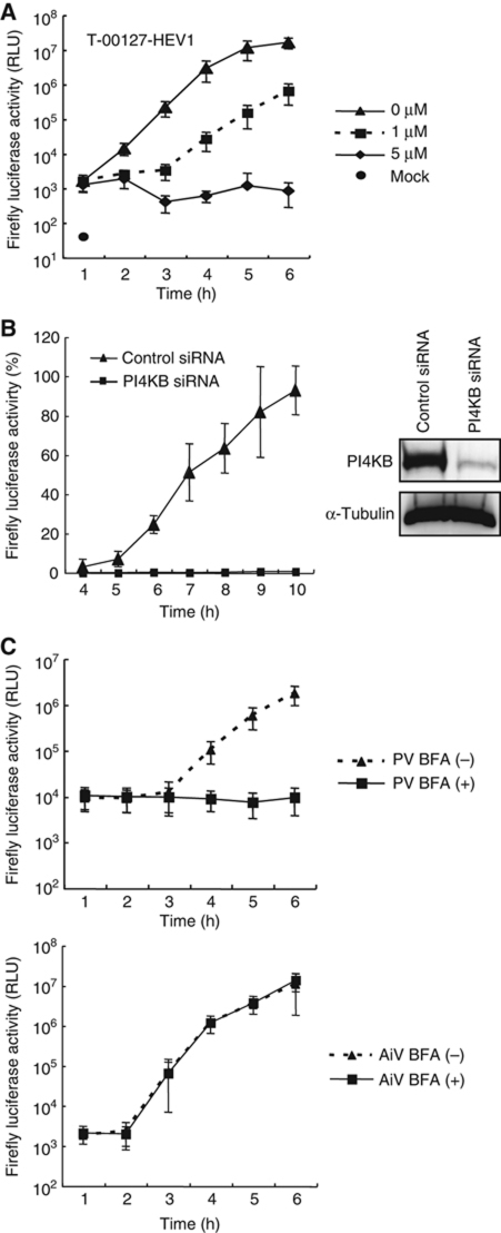
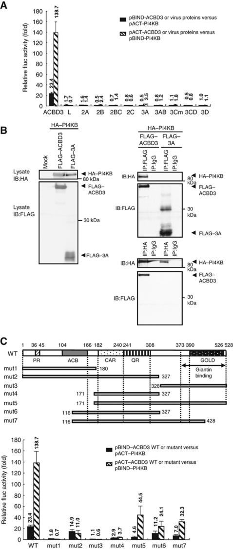
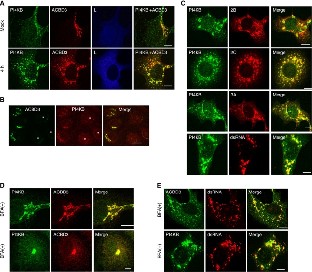
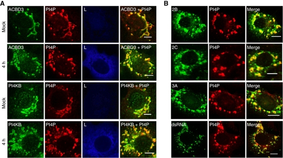
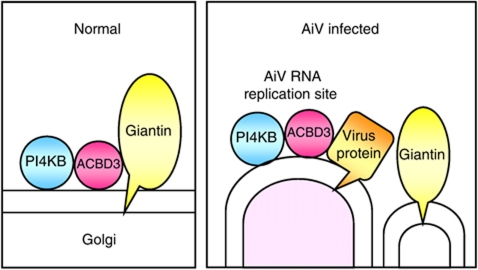
Phosphatidylinositol 4-kinase IIIβ (PI4KB) is a host factor required for genome RNA replication of enteroviruses, small non-enveloped viruses belonging to the family Picornaviridae. Here, we demonstrated that PI4KB is also essential for genome replication of another picornavirus, Aichi virus (AiV), but is recruited to the genome replication sites by a different strategy from that utilized by enteroviruses. AiV non-structural proteins, 2B, 2BC, 2C, 3A, and 3AB, interacted with a Golgi protein, acyl-coenzyme A binding domain containing 3 (ACBD3). Furthermore, we identified previously unknown interaction between ACBD3 and PI4KB, which provides a novel manner of Golgi recruitment of PI4KB. Knockdown of ACBD3 or PI4KB suppressed AiV RNA replication. The viral proteins, ACBD3, PI4KB, and phophatidylinositol-4-phosphate (PI4P) localized to the viral RNA replication sites. AiV replication and recruitment of PI4KB to the RNA replication sites were not affected by brefeldin A, in contrast to those in enterovirus infection. These results indicate that a viral protein/ACBD3/PI4KB complex is formed to synthesize PI4P at the AiV RNA replication sites and plays an essential role in viral RNA replication.


Conflict of interest statement

The authors declare that they have no conflict of interest.

Figures

Figure 1
Figure 1
2B, 2BC, 2C, 3A, and 3AB interact with ACBD3. (A) The mammalian two-hybrid assay. The indicated combination of a pACT construct and a pBIND construct was transfected into Vero cells together with pG5luc encoding a firefly luciferase. Cell lysates were prepared at 48 h after transfection and assayed for firefly luciferase activity. Transfection efficiency was normalized by the activity of Renilla luciferase, which was simultaneously expressed from pBIND. The higher value of normalized luciferase activities obtained in cells transfected with the combination of the pBIND construct and empty pACT and with the combination of the pACT construct and empty pBIND was used as a negative control. The normalized firefly luciferase activity was represented as fold activation compared with a negative control. The experiment was repeated at least three times. Standard deviation bars are shown. (B) Co-immunoprecipitation of ACBD3 with 2B, 2BC, 2C, 3A, or 3AB. FLAG-tagged ACBD3 was co-expressed with HA-tagged L, 2B, 2BC, 2C, 3A, or 3AB. Proteins were immunoprecipitated with anti-FLAG (upper panel), anti-HA (lower panel) antibodies, or control IgG, and the resulting immunoprecipitates and whole cell lysates were analysed by immunoblotting with anti-FLAG and anti-HA antibodies. IB, immunoblotting; IP, immunoprecipitation. (C) MBP pull-down assay. MBP-fused viral proteins or MBP immobilized on amylose resin were mixed with purified GST–ACBD3, and proteins binding to the resin were analysed by SDS–PAGE, followed by immunoblotting with anti-GST antibody (upper panel). After immunoblotting, proteins on a PVDF membrane were stained with Coomassie brilliant blue to detect MBP-fused viral proteins or MBP (lower panel). Figure source data can be found in Supplementary data.
Figure 2
Figure 2
The C-terminal region of ACBD3 interacts with 2B, 2BC, 2C, 3A, and 3AB. (A, C) Schematic representation of (A) full-length ACBD3 (WT) and its mutants (mut1–mut5) and (C) mut3 (amino acids 328–528) and its mutants (mut3Δ1–Δ5). ACBD3 contains characteristic domains as follows: PR, proline-rich domain; ACB, ACB region; CAR, charged amino acid-rich domain; QR, glutamine-rich domain; and GOLD, Golgi dynamic domain. Numbers indicate amino-acid positions. The region between amino acids 373 and 528 is required for binding giantin. (B, D) Mammalian two-hybrid analyses were performed to examine interactions (B) between the ACBD3 mutants (mut1–5) and 2B, 2BC, 2C, 3A, or 3AB, and (D) between the mut3 mutants (mut3Δ1–Δ5) and 2B, 2BC, 2C, 3A, or 3AB, and the results are represented as described in Figure 1A. All experiments were repeated at least three times. Standard deviation bars are shown.
Figure 3
Figure 3
(A) Colocalization of ACBD3 with 2B, 2C, and 3A. Vero cells were electroporated with replicon RNA, AV-FL-Luc-5′rzm. At 4 or 6 h after electroporation, the cells were fixed and double stained with rabbit anti-ACBD3 and guinea pig anti-3A, anti-2B, or anti-2C antibodies. (B) Colocalization of dsRNA with ACBD3, 2B, 2C, or 3A. Vero cells were electroporated with replicon RNA. At 4 h, the cells were fixed and double stained with anti-dsRNA (mouse) and anti-2B, anti-2C, anti-3A, or anti-ACBD3 antibodies (rabbit). Bars represent 30 μm. (C) Effect of knockdown of ACBD3 on AiV replication. Vero cells were transfected with 80 nM of either control siRNA or siRNA against ACBD3. At 72 h after transfection, lysates were prepared and subjected to immunoblotting to assess the levels of ACBD3 and α-tubulin (left panel). At 72 h post transfection with siRNAs, the cells were transfected with replicon RNA and then luciferase activity in cell lysates harvested at the indicated times was measured (right panel). The maximum value obtained for cells treated with control siRNA was taken as 100%. The experiment was repeated at least three times. Standard deviation bars are shown.
Figure 4
Figure 4
Dynamics of different Golgi proteins during AiV RNA replication. Vero cells were mock electroporated or electroporated with the replicon RNA, AV-FL-Luc-5′rzm. At 2 or 4 h after electroporation, the cells were fixed and triple stained with rabbit anti-ACBD3, guinea pig anti-L, and (A) mouse anti-giantin, (B) mouse anti-GM130, or (C) sheep anti-TGN46 antibodies. Bars represent 30 μm.
Figure 5
Figure 5
(A) T-00127-HEV1 inhibits AiV RNA replication. Vero cells were mock electroporated or electroporated with the replicon RNA, AV-FL-Luc-5′rzm, and then treated with 0 (DMSO), 1 or 5 μM T-00127-HEV1. After incubation for the indicated times, the cells were assayed for luciferase activity. (B) Depletion of endogenous PI4KB severely reduces AiV RNA replication. Vero cells were transfected with 40 nM control siRNA or siRNA against PI4KB. At 72 h after transfection, the levels of PI4KB and α-tubulin were determined by immunoblotting (right panel), and the subsequent analysis was performed as in Figure 3C (left panel). (C) AiV RNA replication is insensitive to BFA. Vero cells were electroporated with poliovirus (PV) (upper panel) or AiV (lower panel) replicon RNA and then incubated with or without 10 μg/ml of BFA. After incubation for the indicated times, the cells were assayed for luciferase activity. All experiments were repeated at least three times. Standard deviation bars are shown.
Figure 6
Figure 6
PI4KB interacts with ACBD3. (A) The mammalian two-hybrid assay was performed to examine interactions between PI4KB and ACBD3 or the virus proteins and the results are represented as in Figure 1A. (B) Co-immunoprecipitation of PI4KB with ACBD3. HA-tagged PI4KB was co-expressed with FLAG-tagged ACBD3 or FLAG-tagged 3A in 293T cells, and the subsequent analysis was performed as in Figure 1B. (C) Mapping of the PI4KB-binding region of ACBD3. Schematic representation of full-length ACBD3 (WT) and its mutants (mut6 and mut7) (upper panel). Mammalian two-hybrid analysis was performed to examine interactions between PI4KB and ACBD3 WT or its mutants (mut1–7), and the results are represented as described in Figure 1A (lower panel). Figure source data can be found in Supplementary data.
Figure 7
Figure 7
PI4KB is recruited to the AiV RNA replication sites through interaction with ACBD3 associated with the viral proteins. (A) Colocalization of PI4KB with ACBD3. Vero cells were mock electroporated or electroporated with the replicon RNA, AV-FL-Luc-5′rzm. At 4 h after electroporation, the cells were fixed and triple stained with mouse anti-PI4KB, rabbit anti-ACBD3, and guinea pig anti-L antibodies. (B) Effect of ACBD3 knockdown on the localization of PI4KB. Vero cells were transfected with siRNA against ACBD3. At 72 h after transfection, cells were fixed and double stained with anti-ACBD3 and anti-PI4KB. Asterisks indicate cells where knockdown of the expression of ACBD3 was observed. (C) PI4KB colocalizes with 2B, 2C, 3A, or dsRNA. At 4 h after electroporation with the replicon RNA, the Vero cells were fixed and double stained with mouse anti-PI4KB and rabbit anti-2B, -2C, or -3A antibodies, or rabbit anti-PI4KB and mouse anti-dsRNA antibodies. (D, E) Recruitment of PI4KB and ACBD3 to the AiV RNA replication sites is not affected by BFA. Vero cells were (D) mock electroporated or (E) electroporated with the replicon RNA, and then incubated in medium with or without 10 μg/ml of BFA. At 4 h after electroporation, the cells were fixed and immunostained with the indicated antibodies. Bars represent 30 μm.
Figure 8
Figure 8
PI4P localizes to the AiV RNA replication sites. (A, B) Vero cells were electroporated with mock or replicon RNA. At 4 h after electroporation, the cells were fixed and immunostained with the indicated antibodies. Bars represent 30 μm.
Figure 9
Figure 9
Model for recruitment of PI4KB to the AiV replication sites. In uninfected cells, the C-terminal region of ACBD3 binds to the C-terminal cytoplasmic domain of giantin that is anchored to the Golgi membrane through the C-terminal anchoring domain. PI4KB is localized to the Golgi through interaction with the central part of ACBD3 (left panel). In AiV-infected cells, viral membrane proteins 2B, 2BC, 2C, 3A, and 3AB compete with giantin at the Golgi for binding to ACBD3. The viral protein/ACBD3/PI4KB complex is formed as infection progresses, whereas giantin no longer colocalizes with ACBD3 (right panel).

Similar articles

Cited by

References

    1. Aldabe R, Barco A, Carrasco L (1996) Membrane permeabilization by poliovirus proteins 2B and 2BC. J Biol Chem 271: 23134–23137 - PubMed
    1. Ambert-Balay K, Lorro M, Bon F, Giraudon H, Kaplon J, Wolfer M, Lebon P, Gendrel D, Pothier P (2008) Prevalence and genetic diversity of Aichi virus in community and hospitalized patients. J Clin Microbiol 46: 1252–1258 - PMC - PubMed
    1. Anantharaman V, Aravind L (2002) The GOLD domain, a novel protein module involved in Golgi function and secretion. Genome Biol 3: research0023. - PMC - PubMed
    1. Arita M, Kojima H, Nagano T, Okabe T, Wakita T, Shimizu H (2011) Phosphatidylinositol 4-kinase III beta is a target of enviroxime-like compounds for antipoliovirus activity. J Virol 85: 2364–2372 - PMC - PubMed
    1. Arita M, Nagata N, Sata T, Miyamura T, Shimizu H (2006) Quantitative analysis of poliomyelitis-like paralysis in mice induced by a poliovirus replicon. J Gen Virol 87: 3317–3327 - PubMed

Publication types

Substances