. 2011 Nov;106(6):1069-85.
doi: 10.1007/s00395-011-0231-7. Epub 2011 Nov 29.

Chronic exercise modulates RAS components and improves balance between pro- and anti-inflammatory cytokines in the brain of SHR

Affiliations

Chronic exercise modulates RAS components and improves balance between pro- and anti-inflammatory cytokines in the brain of SHR

Deepmala Agarwal et al. Basic Res Cardiol. 2011 Nov.

Abstract

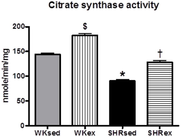
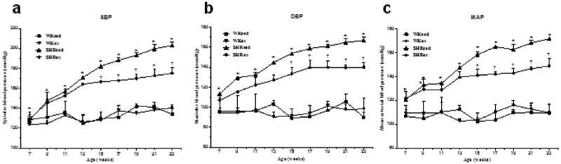
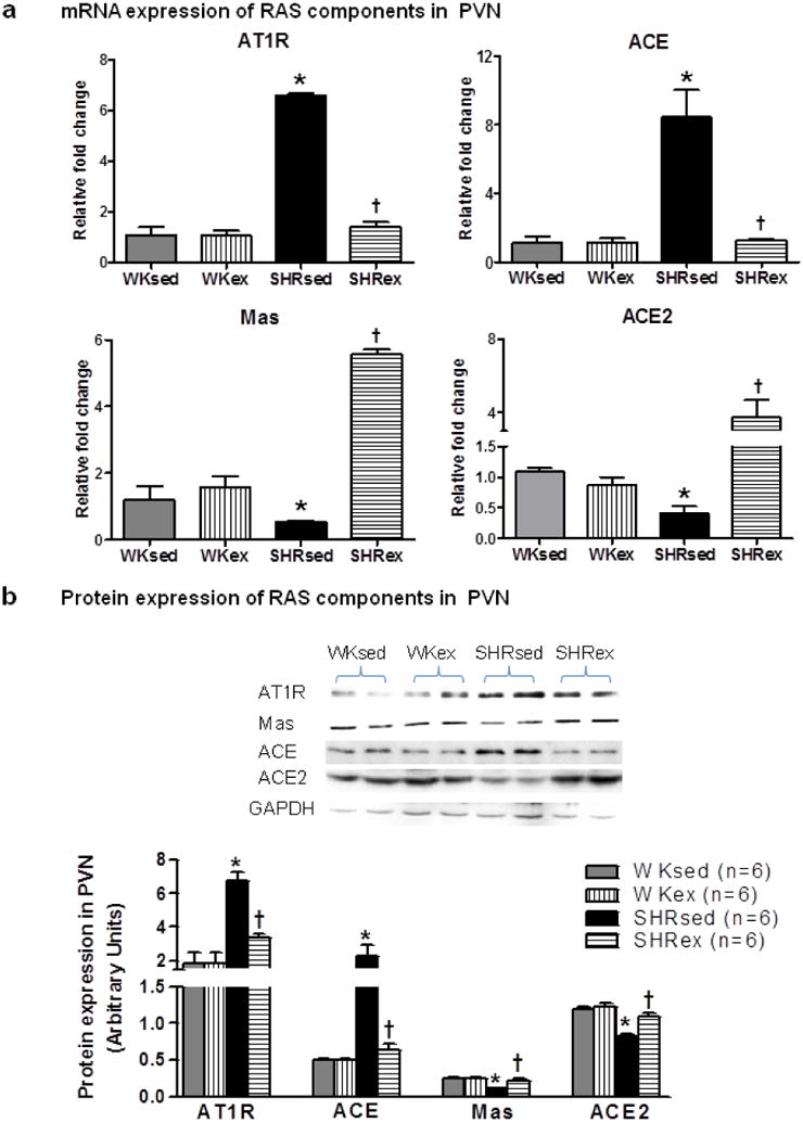
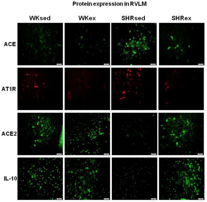
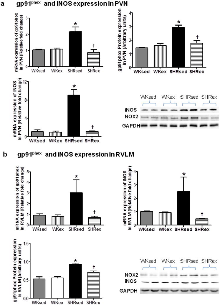
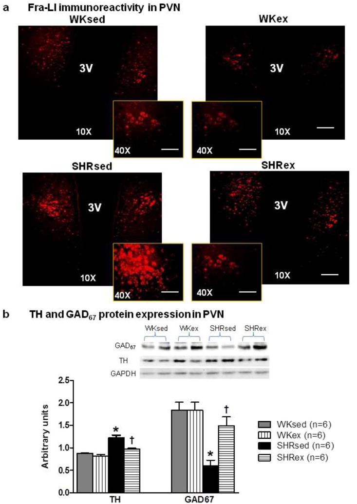
Recently, exercise has been recommended as a part of lifestyle modification for all hypertensive patients; however, the precise mechanisms of its effects on hypertension are largely unknown. Therefore, this study aimed to investigate the mechanisms within the brain that can influence exercise-induced effects in an animal model of human essential hypertension. Young normotensive WKY rats and SHR were given moderate-intensity exercise for 16 weeks. Blood pressure was measured bi-weekly by tail-cuff method. Animals were then euthanized; paraventricular nucleus (PVN) and rostral ventrolateral medulla (RVLM), important cardiovascular regulatory centers in the brain, were collected and analyzed by real-time RT-PCR, Western blot, EIA, and fluorescent microscopy. Exercise of 16-week duration attenuated systolic, diastolic, and mean arterial pressure in SHR. Sedentary SHR exhibited increased pro-inflammatory cytokines (PICs) and decreased anti-inflammatory IL-10 levels in the PVN and RVLM. Furthermore, SHR(sed) rats exhibited elevated levels of ACE, AT1R, and decreased levels of ACE2 and receptor Mas in the PVN and RVLM. Chronic exercise not only prevented the increase in PICs (TNF-α, IL-1β), ACE, and AT1R protein expression in the brain of SHR, but also dramatically upregulated IL-10, ACE2, and Mas receptor expression in SHR. In addition, these changes were associated with reduced plasma AngII levels, reduced neuronal activity, reduced NADPH-oxidase subunit gp91(phox) and inducible NO synthase in trained SHRs indicating reduced oxidative stress. These results suggest that chronic exercise not only attenuates PICs and the vasoconstrictor axis of the RAS but also improves the anti-inflammatory defense mechanisms and vasoprotective axis of the RAS in the brain, which, at least in part, explains the blood pressure-lowering effects of exercise in hypertension.


Conflict of interest statement

Conflict of interest: none declared

Figures

Fig 1
Fig 1
Citrate synthase activity (nmol/min1/mg of protein) in soleus muscle of sedentary or exercised SHR and WKY as measured by citrate synthase activity assay kit. After the period of 16 weeks of exercise, the activity of citrate synthase in the soleus muscle was significantly higher in SHR as well as in WKY rats compared with their sedentary control groups indicating the efficacy of the exercise protocol. $p<0.05 WKsed vs WKex; *p<0.05 WKsed vs SHRsed; †p<0.05 SHRsed vs SHRex
Fig 2
Fig 2
Effect of chronic exercise on time course of blood pressure (in millimeters of mercury) in WK rats and SHRs. A, systolic blood pressure (SBP); B, diastolic blood pressure (DBP); C, mean arterial pressure (MAP). Exercise significantly reduced SBP, DBP, and MAP in SHRex compared with SHRsed rats from 8 weeks of exercise (at 15 weeks of age). Values are mean±SE; n=10 in each group. *p0.05 WKsed vs SHRsed; †p0.05 SHRsed vs SHRex
Fig 3
Fig 3
Effects of exercise on TNF-α, IL-1β, and IL-10 expression in the PVN and RVLM of WK rats and SHRs. mRNA expression in the PVN (a); A representative Western blot (left panel, b) and densitometric analysis (right panel, b) of TNF-α and IL-10 protein expression in the PVN; mRNA expression (c) and densitometric analysis (d) of these cytokines in the RVLM. Values are mean±SE. *p<0.05 WKsed vs. SHRsed; †p<0.05 SHRsed vs SHRex. n=9/group for mRNA analysis and n=6/group for protein analysis
Fig 4
Fig 4
Effects of exercise on RAS components (AT1R, ACE, Mas, and ACE2) in the PVN of WK rats and SHRs. a, mRNA expression; b, densitometric analysis of protein expression and a representative Western blot. Values are mean±SE. *p<0.05 WKsed vs. SHRsed; †p<0.05 SHRsed vs SHRex. n=9/group for mRNA analysis and n=6/group for protein analysis
Fig 5
Fig 5
Effects of exercise on RAS components (AT1R, ACE, Mas, and ACE2) in the RVLM of WK rats and SHRs. a, mRNA expression; b, densitometric analysis of protein expression and a representative Western blot. Values are mean±SE. *p<0.05 WKsed vs. SHRsed; †p<0.05 SHRsed vs SHRex. n=9/group for mRNA analysis and n=6/group for protein analysis
Fig 6
Fig 6
A representative confocal photomicrographs (×20) showing the effects of exercise on protein expression of ACE, ACE2, and TNF-α in the PVN of WK rats and SHRs (n=5/group). Scale bar=100 μ m. 3V,third ventricle
Fig 7
Fig 7
A representative confocal photomicrographs (×20) showing the effects of exercise on protein expression of ACE, AT1R, ACE2, and IL-10 in the RVLM of WK rats and SHRs (n=5/group). Scale bar=100 μm
Fig 8
Fig 8
Effects of exercise on gp91phox and iNOS expression in the PVN and RVLM of WK rats and SHRs. mRNA and protein expression of gp91phox and iNOS in the PVN (a) and RVLM (b). Values are mean±SE. *p<0.05 WKsed vs. SHRsed; †p<0.05 SHRsed vs SHRex. N=9 per group for mRNA analysis and n=6 per group for protein analysis
Fig 9
Fig 9
Effects of exercise on sympathoexcitation in the PVN of WK rats and SHRs. a, A representative confocal photomicrographs (×10, scale bar=200 μ m; and ×0, scale bar=50 μ m) from each group showing Fra-LI immunoreactivity in PVN neurons (n=5/group); b, Densitometric analysis (n=6/group) of protein expression of TH and GAD67 in the PVN accompanied with a representative Western blot. Values are mean±SE; *p<0.05 WKsed vs. SHRsed; †p<0.05 SHRsed vs SHRex. 3V,third ventricle
Fig 10
Fig 10
Densitometric analysis (n=6/group) of protein expression of TH and GAD67 in the RVLM of WK rats and SHRs. In the RVLM, there was no significant difference of TH and GAD67 levels among all groups. Values are mean±SE

References

    1. Abramson JL, Vaccarino V. Relationship between physical activity and inflammation among apparently healthy middle-aged and older US adults. Arch Intern Med. 2002;162:1286–1292. doi: ioi10476. - PubMed
    1. Adams V, Linke A, Krankel N, Erbs S, Gielen S, Mobius-Winkler S, Gummert JF, Mohr FW, Schuler G, Hambrecht R. Impact of regular physical activity on the NAD(P)H oxidase and angiotensin receptor system in patients with coronary artery disease. Circulation. 2005;111:555–562. doi: 10.1161/01.CIR.0000154560.88933.7E. - DOI - PubMed
    1. Agarwal D, Haque M, Sriramula S, Mariappan N, Pariaut R, Francis J. Role of proinflammatory cytokines and redox homeostasis in exercise-induced delayed progression of hypertension in spontaneously hypertensive rats. Hypertension. 2009;54:1393–1400. doi: 10.1161/HYPERTENSIONAHA.109.135459. - DOI - PMC - PubMed
    1. Bai Y, Jabbari B, Ye S, Campese VM, Vaziri ND. Regional expression of NAD(P)H oxidase and superoxide dismutase in the brain of rats with neurogenic hypertension. Am J Nephrol. 2009;29:483–492. doi: 10.1159/000178817. - DOI - PMC - PubMed
    1. Block CH, Santos RA, Brosnihan KB, Ferrario CM. Immunocytochemical localization of angiotensin-(1-7) in the rat forebrain. Peptides. 1988;9:1395–1401. doi: 10.1016/0196-9781(88)90208-2. - DOI - PubMed

Publication types

MeSH terms