Review
. 2012 Apr;3(1-2):14-25.
doi: 10.1007/s12672-011-0097-z.

Mechanisms of HGF/Met signaling to Brk and Sam68 in breast cancer progression

Affiliations
Review

Mechanisms of HGF/Met signaling to Brk and Sam68 in breast cancer progression

Alessia Locatelli et al. Horm Cancer. 2012 Apr.

Abstract

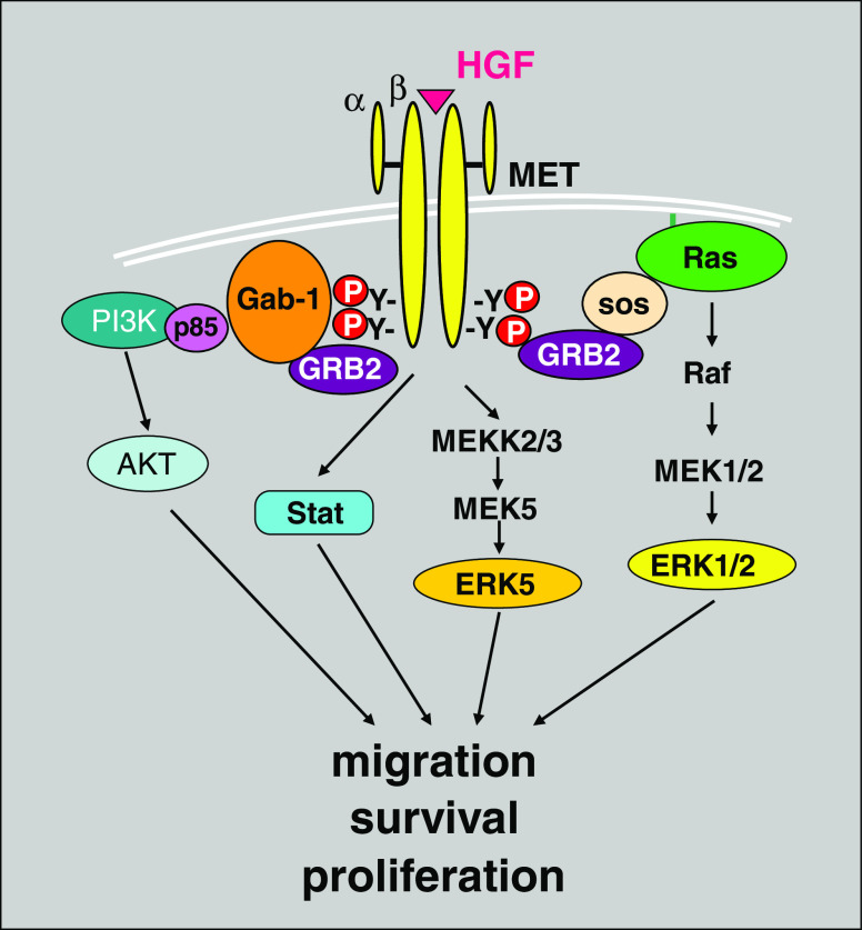
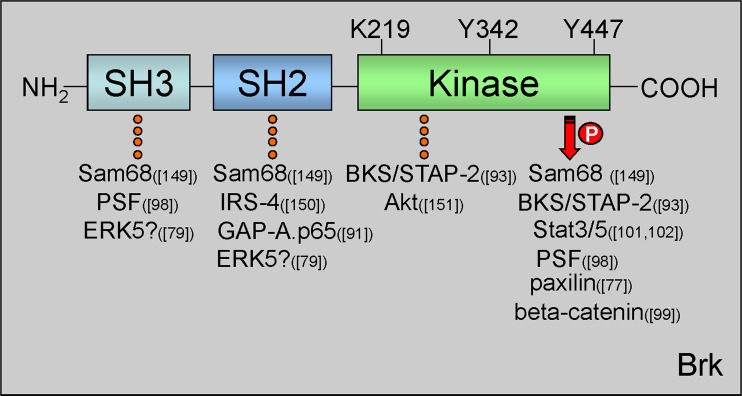
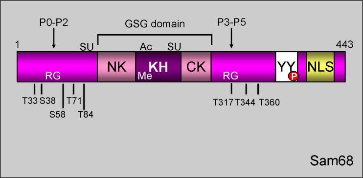
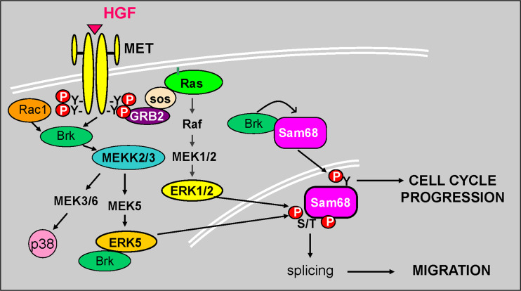
Signal transduction pathways downstream of receptor tyrosine kinases (RTKs) are often deregulated during oncogenesis, tumor progression, and metastasis. In particular, the peptide growth factor hormone, hepatocyte growth factor (HGF), and its specific receptor, Met tyrosine kinase, regulate cancer cell migration, thereby conferring an aggressive phenotype (Nakamura et al., J Clin Invest 106(12):1511-1519, 2000; Huh et al., Proc Natl Acad Sci U S A 101:4477-4482, 2004). Additionally, overexpression of Met is associated with enhanced invasiveness of breast cancer cells (Edakuni et al., Pathol Int 51(3):172-178, 2001; Jin et al., Cancer 79(4):749-760, 1997; Tuck et al., Am J Pathol 148(1):225-232, 1996). Here, we review the regulation of recently identified novel downstream mediators of HGF/Met signaling, Breast tumor kinase (Brk/PTK6), and Src-associated substrate during mitosis of 68 kDa (Sam68), and discuss their relevance to mechanisms of breast cancer progression.


Figures

Fig. 1
Fig. 1
HGF/Met receptor signaling pathway. Stimulation of Met receptor by its specific ligand HGF leads to activation of a variety of downstream signaling molecules, targeting cell proliferation, migration, and survival
Fig. 2
Fig. 2
Brk protein domains and important regulatory residues. Brk protein structure consists of one N-terminal SH3 domain, one SH2 domain, and a C-terminal kinase domain. Within the kinase domain are three residues important for Brk kinase activity. Lys219 (K219) in the ATP binding pocket is required for kinase activation. Mutation of this site to Met (M) renders Brk kinase-inactive. Tyr342 (Y342) is autophosphorylated upon Brk activation. Tyr447 (Y447) is required for Brk autoinhibition. Substitution of Y to Phe (F) at this site mimics phosphorylation and results in a constitutively active Brk molecule. Brk domain-specific interacting proteins are indicated with dotted line and substrates with arrow; references to the specific proteins are reported in parenthesis [, , , , , , , , –150]. PTB polypyrymidine tract, PSF protein-associated splicing factor, IRS-4 insulin receptor substrate 4, BKS/STAP2 breast tumor kinase substrate
Fig. 3
Fig. 3
Schematic representation of Sam68 structural/functional domains. Sam68 is composed of a GSG domain, consisting of a single RNA-binding KH domain flanked by NK (N-terminal of KH) and CK (C-terminal of KH) segments, six consensus proline-rich motifs (P0–P5), arginine-rich boxes (RG), C-terminal tyrosine-rich domain (YY), and an NLS. Sam68 undergoes post-translational modification such as SUMOylation (SU), acetylation (Ac), methylation (Me), and phosphorylation (P)
Fig. 4
Fig. 4
Mechanism of action of Brk and Sam68 downstream of activated Met receptors. Brk and Sam68 are essential effectors of HGF-induced cell cycle progression (Brk-mediated Tyr phosphorylation of Sam68) and migration (MAPK-mediated Ser/Thr phosphorylation of Sam68) via an ERK5-dependent mechanism. Growth factor activation facilitates Brk complex formation with ERK5 activating both tyrosine and serine/threonine phosphorylation events which lead to downstream biological outcomes. Brk also associates with and phosphorylates Sam68 to alter its RNA binding activity and thus cell cycle progression. HGF binding to Met receptors induces Brk, Erk5, and Sam68 dependent cellular migration
Fig. 5
Fig. 5
EGFR activity is required for HGF-induced phosphorylation of Met receptors and activation of Erk5. MDA-MB-231 breast cancer cells were pretreated with AG1478 (EGFR tyrosine kinase inhibitor) for 30 min prior to HGF stimulation for 15 min. Cells were harvested and Western blotted with antibodies against phospho-Met receptor, total Met receptor, and total Erk5. The up-shifted higher migrating band in the total Erk5 blot represents phosphorylated (i.e., on multiple sites) and activated Erk5

Similar articles

Cited by

References

    1. Nakamura T, et al. Myocardial protection from ischemia/reperfusion injury by endogenous and exogenous HGF. J Clin Invest. 2000;106(12):1511–1519. - PMC - PubMed
    1. Huh CG, et al. Hepatocyte growth factor/c-met signaling pathway is required for efficient liver regeneration and repair. Proc Natl Acad Sci U S A. 2004;101:4477–4482. - PMC - PubMed
    1. Edakuni G, et al. Expression of the hepatocyte growth factor/c-Met pathway is increased at the cancer front in breast carcinoma. Pathol Int. 2001;51(3):172–178. - PubMed
    1. Jin L, et al. Expression of scatter factor and c-met receptor in benign and malignant breast tissue. Cancer. 1997;79(4):749–760. - PubMed
    1. Tuck AB, et al. Coexpression of hepatocyte growth factor and receptor (Met) in human breast carcinoma. Am J Pathol. 1996;148(1):225–232. - PMC - PubMed

Publication types

MeSH terms