. 2012 Jan;9(1):210-25.
doi: 10.1007/s13311-011-0084-7.

Glucocorticoid-induced leucine zipper (GILZ) over-expression in T lymphocytes inhibits inflammation and tissue damage in spinal cord injury

Affiliations

Glucocorticoid-induced leucine zipper (GILZ) over-expression in T lymphocytes inhibits inflammation and tissue damage in spinal cord injury

Emanuela Esposito et al. Neurotherapeutics. 2012 Jan.

Abstract

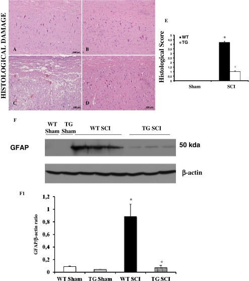
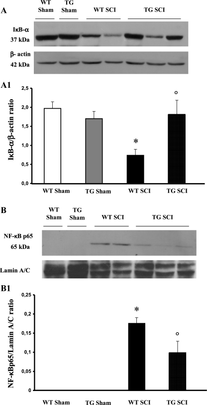
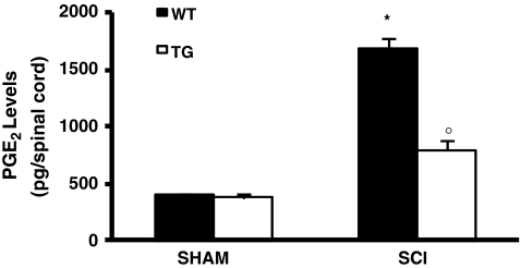
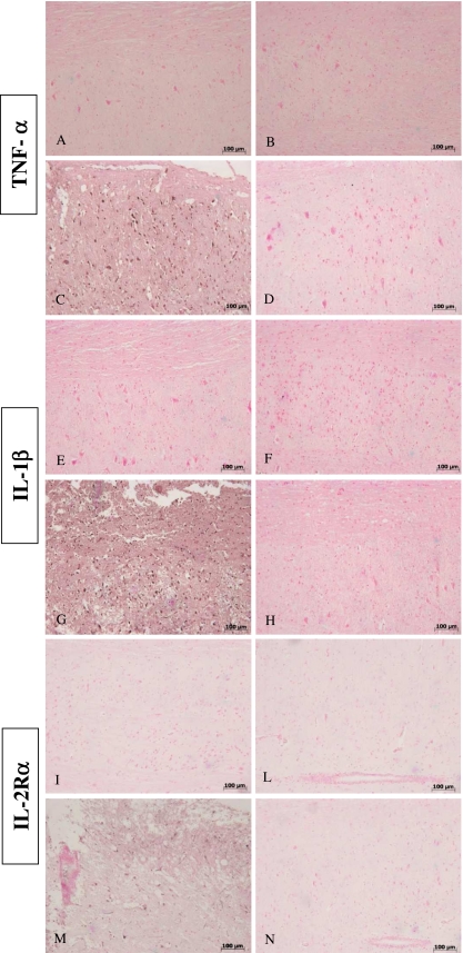
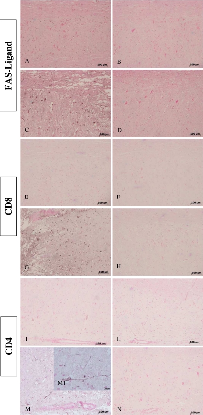
Spinal cord injury (SCI) is a traumatic event that causes a secondary and extended inflammation characterized by infiltration of immune cells, including T lymphocytes, release of pro-inflammatory mediators in the lesion site, and tissue degeneration. Current therapeutic approaches for SCI are limited to glucocorticoids (GC) due to their potent anti-inflammatory activity. GC efficacy resides, in part, in the capability to inhibit NF-κB, T lymphocyte activation, and the consequent cytokine production. In this study, we performed experiments aimed to test the susceptibility of glucocorticoid-induced leucine zipper (GILZ) transgenic (GILZ(TG)) mice, in which GILZ is selectively over-expressed in T lymphocytes, to SCI induction. Consistent with a decreased inflammatory response, GILZ(TG) were less susceptible to SCI as compared to wild-type littermates. Notably, inhibition of NF-κB activation and nuclear translocation, diminished T lymphocytes activation and tissue infiltration, as well as decreased release of cytokines were evident in GILZ(TG) as compared to wild-type mice. Moreover, GILZ(TG) showed a reduced tumor necrosis factor-α, IL-1β, Inductible nitric oxide synthase (iNOS) and nytrotyrosine production, apoptosis, and neuronal tissue damage. Together these results indicate that GILZ mimics the anti-inflammatory effect of GC and represents a potential pharmacological target for modulation of T lymphocyte-mediated immune response in inflammatory disorders, such as SCI.


Figures

Fig. 1
Fig. 1
Effect of GILZ on histological alterations of the spinal cord tissue and immunohistochemical localization of iNOS and nitrotyrosine. No histological alteration was observed in the spinal cord tissues from sham-operated mice (A,B). Twenty-four h after SCI a significant damage was evident in spinal cord from WT mice, as also indicated by the presence of edema as well as alteration of the white matter (C). Notably, a significant protection from the SCI was observed in the tissue from GILZTG mice (D). The histological scoring of injury severity showed that the degree of injury is higher in mice SCI-operated mice, when compared with SCI GILZTG mice and sham-operated mice (E). Moreover, the cellular changes occurring in the perilesioned zone (namely, at the boundary between the core necrotic area and the penumbra zone) were evaluated by the expression of GFAP, a specific glial/precursor cell markers. The increased levels of GFAP protein were significantly reduced in the spinal cord tissues from GILZTG as compared to WT mice (F,F1). Immunoblottings in panel F is representative of one spinal cord out of 5 analyzed. The densitometry results in panel F1 are expressed as mean ± s.e.m. of 3 blots. Histological score data are expressed as mean ± s.e.m. of 6 mice each group. *p < 0.01 vs. sham; °p < 0.01 vs. SCI-operated mice. GILZ over-expression in SCI operated mice produced a marked reduction in the immunostaining for iNOS (L) and nitrotyrosine (P) in spinal cord tissue, when compared to positive iNOS (I) and nitrotyrosine (O) staining obtained from the spinal cord tissue from WT mice 24 h after the injury. No positive staining was detected in sham-operated mice for iNOS or nitrotyrosine (G, H and M,N respectively). This figure is representative of 3 experiments performed on different experimental days
Fig. 2
Fig. 2
Effect of GILZ over-expression on IκB-a degradation and NF-κB p65 nuclear translocation. A basal level of IκB-a was detected in the spinal cord from sham-operated animals (A, A1) that was substantially reduced 24 h after SCI induction in WT mice (A, A1). GILZ over-expression, in GILZTG mice, prevented the SCI-induced IκB-a reduction (A, A1). In addition, the levels of NF-κB p65 protein were significantly reduced in the nuclear fractions of the spinal cord tissues animals from GILZTG as compared to WT mice (B,B1). Immunoblottings in panel A and B are representative of one spinal cord out of 5 analyzed. The densitometry results in panel A1 and B1 were obtained by phosphoimager analysis and are expressed as mean ± s.e.m. of 2 blots *p < 0.05 versus sham, °p < 0.05 versus SCI-operated mice
Fig. 3
Fig. 3
Effect of GILZ over-expression on MPO activity, production of TNF-a, IL-1b, and IL-10. Myeloperoxidase (MPO) levels were increased by spinal cord injury in WT mice as compared with tissues obtained from sham animals. In contrast, a decrease MPO activity was observed in tissue taken from GILZTG mice 24 h after injury (A). The evaluation of the production of inflammatory cytokines IL-10, TNF-a and IL-1b, by ELISA, showed that in spinal cord samples, taken after 24 h after injury, there was a substantial increase in IL-10 (B), TNF-a (C) and IL-1b (D) formation when compared with sham-operated animals. In contrast, in GILZTG mice there were a significant increase of IL-10 (B), and inhibition of TNF-a (C) and IL-1b (D) as compared to WT. Data are mean ± s.e.m. of 6 mice for each group. *p < 0.01 vs sham, °p < 0.01 vs SCI-operated mice
Fig. 4
Fig. 4
Effect of GILZ over-expression on PGE2 levels PGE2 levels were increased by SCI in WT mice as compared with tissues obtained from sham animals. Mice with GILZ over-expression showed a reduction of PGE2 levels
Fig. 5
Fig. 5
Effects of GILZ on tissue expression of TNF-a, IL-1b and IL-2Ra, after SCI, as evaluated by immuno-histochemistry Positive staining for TNF-a (C) and IL-1b (G) was observed in spinal cord tissue sections obtained from SCI GILZWT animals. The staining for TNF-a (D) and IL-1b (H) in SCI GILZTG mice was reduced significantly in the tissues collected at 24 h. Spinal cord sections from sham-operated mice not shown stain for TNF-a (A,B) and IL-1b (E,F) Moreover, spinal cord sections from sham-operated mice did not stain for IL-2Ra (I and L). Spinal cord sections obtained from WT mice exhibited positive staining for IL-2Ra (M) after SCI induction. GILZ over-expression in GILZTG mice reduced the degree of positive staining for IL-2Ra in the spinal cord (N)
Fig. 6
Fig. 6
Effects of GILZ on apoptosis as evaluated by TUNEL coloration in spinal cord tissue sections. The number of apoptotic fragments and cells increased at 24 h after SCI induction (C), associated with a specific apoptotic morphology characterized by the compaction of chromatin into uniformly dense masses in perinuclear membrane, the formation of apoptotic bodies as well as the membrane blebbing (see particle C1). In contrast, tissues obtained from GILZTG mice (D) demonstrated a small number of apoptotic cells or fragments. No staining was found in shamoperated mice (A-B). In SHAM-operated animals, the central area of each tissue section was taken as a reference point and an identical number of optical fields was counted (see Supplementary Figure 1 for details). Data are reported as mean ± standard error. For each staining, at least 20 optical fields from sections taken from 3 different animals were counted. *p < 0.01 vs. sham; °p < 0.01 vs. SCI-operated mice
Fig. 7
Fig. 7
Effects of GILZ on Bax and Bcl-2 levels, as evaluated by western blot and by immunohistochemical staining. Western blot analysis was realized in spinal cord tissue collected at 24 h after injury. (A) Sham: basal level of Bax was present in the tissue from-sham-operated mice. SCI WT: Bax band is more evident in the tissue from spinal cord injured mice. SCI GILZTG: Bax band is reduced in the tissue from spinal cord injured mice. (B) Sham: basal level of Bcl-2 was present in the tissue from-sham-operated mice. SCI WT: Bcl-2 band is reduced in the tissue from spinal cord injured mice. SCI GILZTG: Bcl-2 band is more evident in the tissue from spinal cord injured mice. Immunoblottings in panel A and B are representative of one spinal cord tissues out of 3 analyzed. The results in panel A1 and B1 (densitometry measured by Phosphoimage analysis, GE Healthcare, Milan, Italy) are expressed as mean ± s.e.m.. *p < 0.01 vs. Sham; °p < 0.01 vs. SCI GILZWT. No positive staining for Bax was observed in the tissue section from sham-operated mice (C,D). SCI caused, at 24 h, an increase in the release of Bax expression (E). GILZ over-expression significantly inhibited the SCI-induced increase in Bax expression (F). On the contrary positive staining for Bcl-2 was observed in the spinal cord tissues of sham-operated mice (G,H). At 24h after SCI significantly less staining for Bcl-2 was observed (I). GILZ over-expression significantly prevented the loss of Bcl-2 expression induced by SCI (L). Figure is representative of at least 3 experiments performed on different experimental days
Fig. 8
Fig. 8
Effects of GILZ on expression of FasL, CD4+ and CD8a+ T cells, after SCI. Immunohistological staining for FasL in the spinal cord was also determined 24 h after injury. No positive staining for sham-operated mice (A, B). Spinal cord sections obtained from SCI WT mice exhibited positive staining (C) mainly localized in inflammatory cells as well as in nuclei of Schwann cells. Inhibition of expression of GILZ reduced the degree of positive staining for FasL in the spinal cord (D). Spinal cords obtained from SCI WT mice showed significant accumulation of CD8a+ (G) and CD4+ (M, particle M1) T cells, when compared with sham operated animals (E,F and I,L, respectively). GILZ over-expression reduced the degree of positive staining for CD8a+ (H) and CD4+ (N) T cells in the spinal cord.

Similar articles

Cited by

References

    1. Malmberg AB, Yaksh TL. Cyclooxygenase inhibition and the spinal release of prostaglandin E2 and amino acids evoked by paw formalin injection: a microdialysis study in unanesthetized rats. J Neurosci. 1995;15:2768–2776. - PMC - PubMed
    1. Bartholdi D, Schwab ME. Methylprednisolone inhibits early inflammatory processes but not ischemic cell death after experimental spinal cord lesion in the rat. Brain Res. 1995;672:177–186. doi: 10.1016/0006-8993(94)01410-J. - DOI - PubMed
    1. Amar AP, Levy ML. Pathogenesis and pharmacological strategies for mitigating secondary damage in acute spinal cord injury. Neurosurgery. 1999;44:1027–1040. doi: 10.1097/00006123-199905000-00052. - DOI - PubMed
    1. Carlson SL, Parrish ME, Springer JE, Doty K, Dossett L. Acute inflammatory response in spinal cord following impact injury. Exp Neurol. 1998;151:77–88. doi: 10.1006/exnr.1998.6785. - DOI - PubMed
    1. Castro R, Jr HMG, Xu GY, et al. Evidence that infiltrating neutrophils do not release reactive oxygen species in the site of spinal cord injury. Exp Neurol. 2004;190:414–424. doi: 10.1016/j.expneurol.2004.05.046. - DOI - PubMed

Publication types

MeSH terms