

Activation likelihood estimation meta-analysis of brain correlates of placebo analgesia in human experimental pain
- PMID: 22125184
- PMCID: PMC6870130
- DOI: 10.1002/hbm.21471
Activation likelihood estimation meta-analysis of brain correlates of placebo analgesia in human experimental pain
Abstract
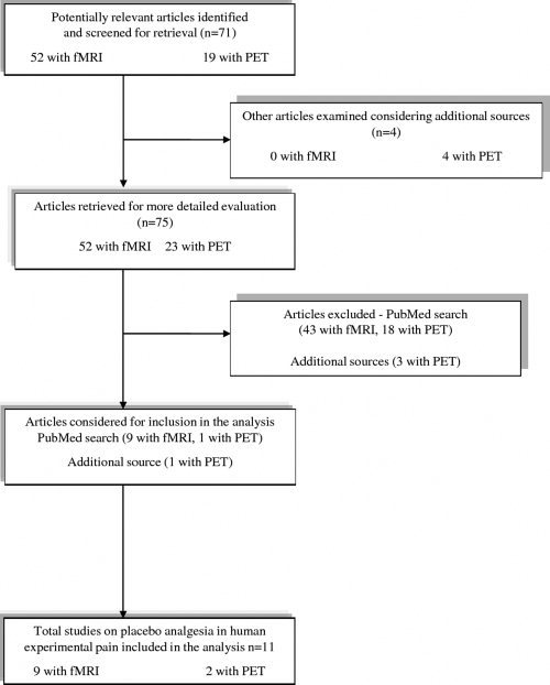
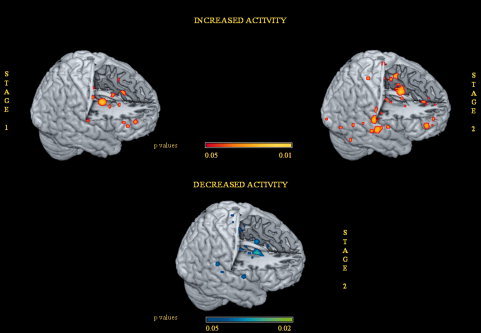
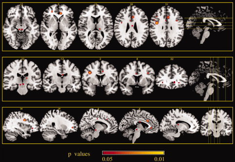
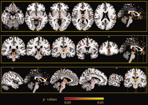
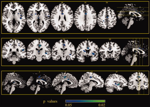
Placebo analgesia (PA) is one of the most studied placebo effects. Brain imaging studies published over the last decade, using either positron emission tomography (PET) or functional magnetic resonance imaging (fMRI), suggest that multiple brain regions may play a pivotal role in this process. However, there continues to be much debate as to which areas consistently contribute to placebo analgesia-related networks. In the present study, we used activation likelihood estimation (ALE) meta-analysis, a state-of-the-art approach, to search for the cortical areas involved in PA in human experimental pain models. Nine fMRI studies and two PET studies investigating cerebral hemodynamic changes were included in the analysis. During expectation of analgesia, activated foci were found in the left anterior cingulate, right precentral, and lateral prefrontal cortex and in the left periaqueductal gray (PAG). During noxious stimulation, placebo-related activations were detected in the anterior cingulate and medial and lateral prefrontal cortices, in the left inferior parietal lobule and postcentral gyrus, anterior insula, thalamus, hypothalamus, PAG, and pons; deactivations were found in the left mid- and posterior cingulate cortex, superior temporal and precentral gyri, in the left anterior and right posterior insula, in the claustrum and putamen, and in the right thalamus and caudate body. Our results suggest on one hand that the modulatory cortical networks involved in PA largely overlap those involved in the regulation of emotional processes, on the other that brain nociceptive networks are downregulated in parallel with behavioral analgesia.
Copyright © 2011 Wiley Periodicals, Inc.
Figures
References
-
- Amanzio M, Torta DME, Sacco K, Cauda F, D'Agata F, Duca S, Leotta D, Palermo S, Geminiani G ( 2011): Unawareness of deficits in Alzheimer's disease: Role of the cingulate cortex. Brain 134: 1061–1076. - PubMed
-
- Bechara A, Damasio H, Tranel D, Damasio AR ( 1997): Deciding advantageously before knowing the advantageous strategy. Science 275: 1293–1295. - PubMed
-
- Benedetti F ( 1996): The opposite effects of the opiate antagonist naloxone and the cholecystokinin antagonist proglumide on placebo analgesia. Pain 64: 535–543. - PubMed
