. 2012 Jan 17;53(1):143-9.
doi: 10.1167/iovs.11-8174.

The accommodative lag of the young hyperopic patient

Affiliations

The accommodative lag of the young hyperopic patient

T Rowan Candy et al. Invest Ophthalmol Vis Sci. .

Abstract

Purpose: To determine the accommodative accuracy of infants and young children before they had had any form of clinical intervention or treatment, in an attempt to determine the difference between 'normal' and 'abnormal' visual experience for these individuals.

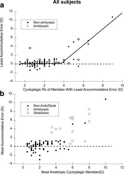
Methods: Nott retinoscopy was performed on 111 subjects in binocular viewing conditions at a viewing distance of 50 cm. The target was a naturalistic cartoon image with a broadband spatial frequency amplitude spectrum.

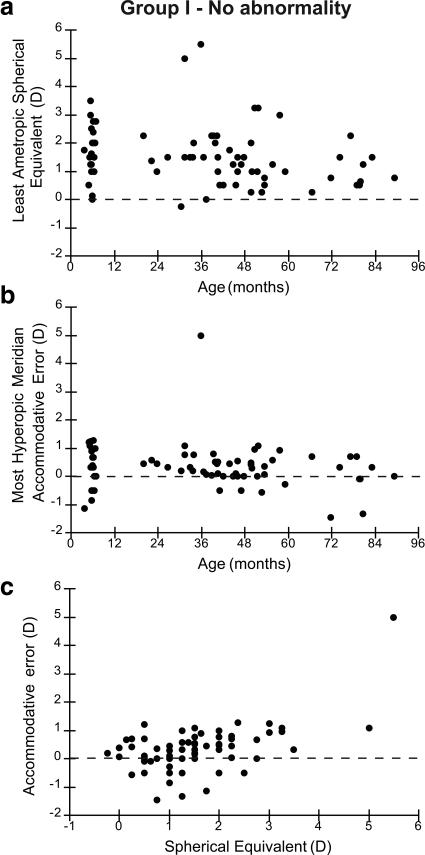
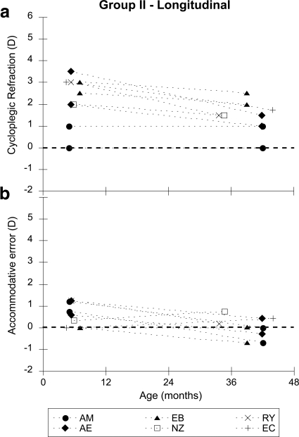
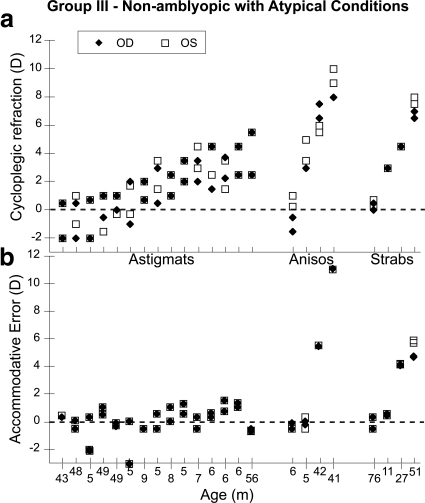
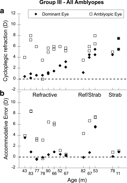
Results: Accommodative accuracy was not related to age (4-90 months). In the group found to have no apparent clinical abnormality (n = 71), the mean lag in the more hyperopic meridian of the least ametropic eye was 0.34 diopters (D). When considering the group as a whole, those with less than approximately 4 D of hyperopia demonstrated similar lags, while those with higher hyperopia, amblyopia, or strabismus had more variable lags. An ROC analysis designed to detect hyperopia >5 D in any meridian, amblyopia and/or strabismus had an area under the curve of 0.90 (95% confidence interval [CI], 0.82 to 0.95), and for a lag criterion of 1.3 D had a sensitivity of 83.3% and a specificity of 96.5%.

Conclusions: These data from a relatively small but broad sampling of age and clinical status suggest that clinically normal young infants and children with low amounts of hyperopia have similar lags of accommodation from the first few months after birth. Subjects with greater than 4 D of hyperopia, or amblyopia or strabismus, have more variable lags and therefore evidence of abnormal visual experience.


Figures

Figure 1.
Figure 1.
Data from group I, who had no clinical abnormality as defined by the inclusion criteria described in the text. (a) Spherical equivalent cycloplegic refractive error of the least ametropic eye is plotted as a function of age. (b) The accommodative error of the most hyperopic meridian of the least ametropic eye is plotted as a function of age. (c) Accommodative error of most hyperopic meridian is plotted as a function of spherical equivalent refractive error of the least ametropic eye.
Figure 2.
Figure 2.
Data from group II, who were tested at both 6 months and 2 or 3 years of age. Each symbol represents a different subject and is labeled with their initials. The filled symbols represent subjects with astigmatism (two principal meridia). (a) The refractive error of the principal meridia of their least hyperopic eye is plotted as a function of age. (b) The accommodative error of those meridia is plotted as a function of age.
Figure 3.
Figure 3.
Data from the nonamblyopic subjects in group III, who all had astigmatism, anisometropia, or strabismus. The subjects are ordered based on increasing refractive error, with the subject group labeled on the abscissa of (a) and age labeled on the abscissa of (b). (a) Cycloplegic refractive error for all meridia plotted for each subject. (b) Accommodative error for all meridia plotted for the vertically corresponding subject in each group.
Figure 4.
Figure 4.
Data from the amblyopic subjects in group III. The subjects are ordered based on increasing refractive error, with the subject group labeled on the abscissa of (a) and age labeled on the abscissa of (b). (a) Cycloplegic refractive error for all meridia plotted for each subject. (b) Accommodative error for all meridia plotted for the vertically corresponding subject in each group. The fact that the data were collected in binocular viewing means that the more hyperopic eyes of the anisometropics are more defocused merely as a result of the accurate accommodation in the dominant eye.
Figure 5.
Figure 5.
Two analyses of accommodative error of the whole group. (a) Accommodative error for the meridian focused closest to the target in binocular viewing conditions plotted as a function of refractive error in that meridian. The full group of subjects is split into two groups, amblyopes and nonamblyopic subjects. The lines fit to the data are described in the text. (b) Least accurate accommodative error of any meridian plotted as a function of cycloplegic refractive error in the most ametropic meridian from either eye of nonstrabismic amblyopes, strabismics, and the other more typically-developing children with no amblyopia or strabismus.

Similar articles

Cited by

References

    1. Mitchell DE, Timney B. Postnatal development of function in the mammalian visual system. In: Kandel ER, ed. Handbook of Physiology. The Nervous System III. Vol. 3 Washington DC: American Physiological Society; 1984:507–555
    1. Movshon JA, Eggers HM, Gizzi MS, Hendrickson AE, Kiorpes L, Boothe RG. Effects of early unilateral blur on the macaque's visual system. III. Physiological observations. J Neurosci. 1987;7:1340–1351 - PMC - PubMed
    1. Kiorpes L, Kiper DC, O'Keefe LP, Cavanaugh JR, Movshon JA. Neuronal correlates of amblyopia in the visual cortex of macaque monkeys with experimental strabismus and anisometropia. J Neurosci. 1998;18:6411–6424 - PMC - PubMed
    1. Troilo D. Neonatal eye growth and emmetropisation–a literature review. Eye. 1992;6:154–160 - PubMed
    1. Smith EI. Environmentally induced refractive errors in animals. In: Rosenfield M, Gilmartin B, eds. Myopia and Nearwork. Oxford: Butterworth-Heinemann; 1998:57–90

Publication types