

doi: 10.1016/j.progpolymsci.2011.06.003.
Alginate: properties and biomedical applications
Affiliations
- PMID: 22125349
- PMCID: PMC3223967
- DOI: 10.1016/j.progpolymsci.2011.06.003
Item in Clipboard
Alginate: properties and biomedical applications
Prog Polym Sci.
2012 Jan.
Abstract
Alginate is a biomaterial that has found numerous applications in biomedical science and engineering due to its favorable properties, including biocompatibility and ease of gelation. Alginate hydrogels have been particularly attractive in wound healing, drug delivery, and tissue engineering applications to date, as these gels retain structural similarity to the extracellular matrices in tissues and can be manipulated to play several critical roles. This review will provide a comprehensive overview of general properties of alginate and its hydrogels, their biomedical applications, and suggest new perspectives for future studies with these polymers.
Figures
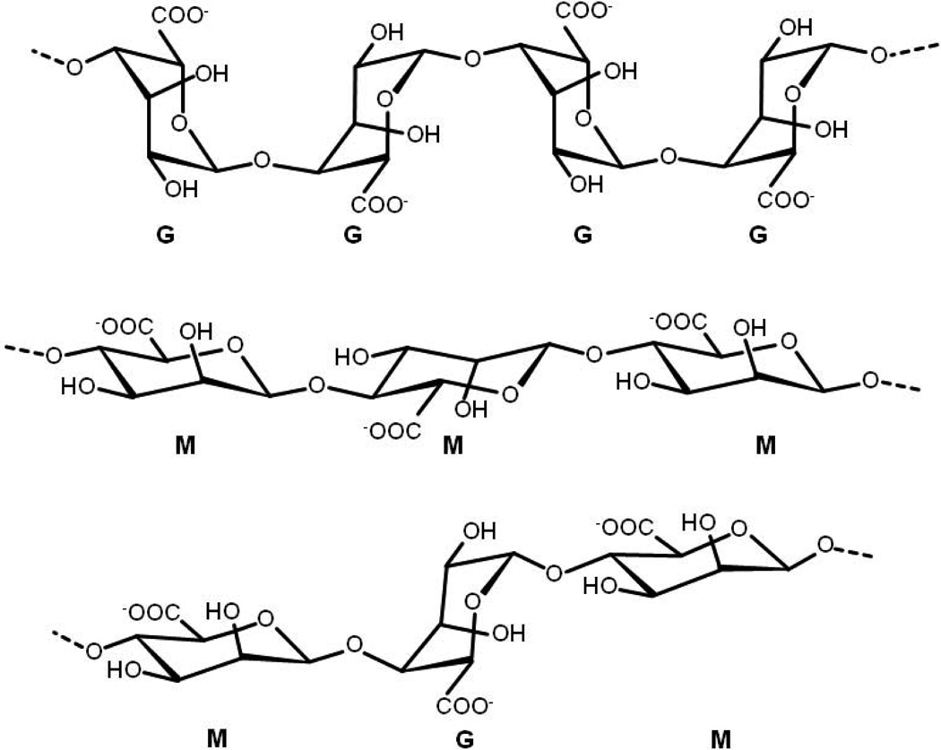
Chemical structures of G-block, M-block, and alternating block in alginate.
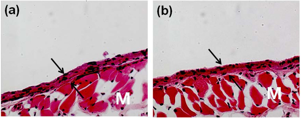
Photomicrographs of tissue sections three weeks post-injection with (a) PBS and (b) alginate hydrogels. Original pictures were taken at 100× magnification. Photomicrographs have labels for the muscle layer (M). Arrows indicate the newly formed granulation tissue adjacent to the site of injection: this is a connective tissue typically formed following biomaterial implantation [24]. Copyright 2009, Springer, New York, USA.
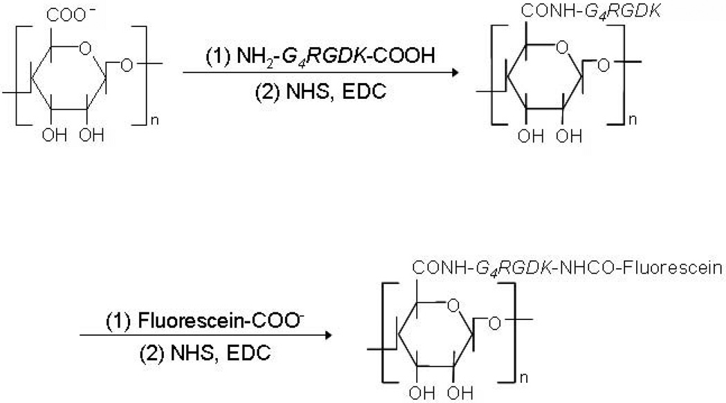
Synthetic scheme of RGD-modified alginate using water-soluble carbodiimide chemistry (NHS, N-hydroxysulfosuccinimide; EDC, 1-ethyl-3-(dimethylaminopropyl) carbodiimide). For further analysis, RGD peptide can be labeled with fluorescein [33], as demonstrated in this scheme. Copyright 2008, WILEY-VCH Verlag GmbH & Co. KGaA, Weinheim, Germany.
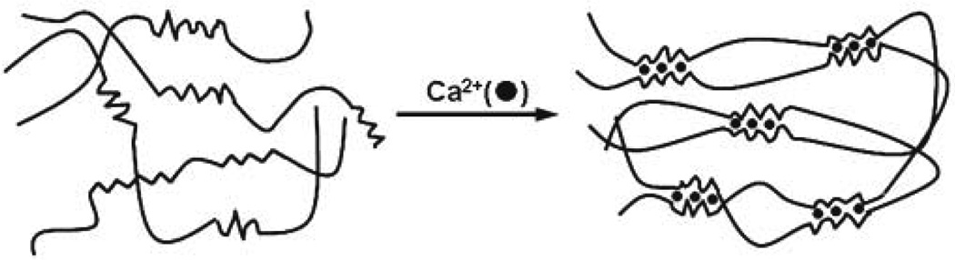
Alginate hydrogels prepared by ionic cross-linking (egg-box model) [38]. Only guluronate blocks participate in the formation of a corrugated egg-box-like structure with interstices in which calcium ions are placed. Copyright 2007, Elsevier Science Ltd., Oxford, UK.
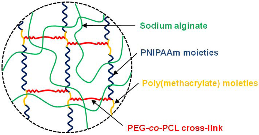
Schematic description of thermo-sensitive semi-IPN alginate hydrogels [54].
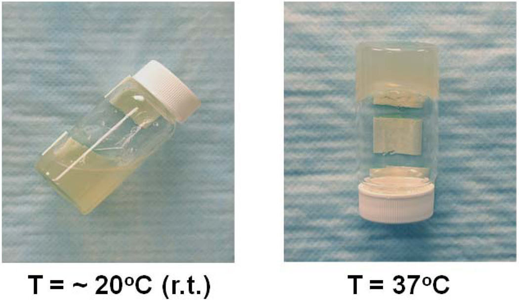
Thermal gelation of an aqueous alginate-g-NIPAAm solution at 37°C.
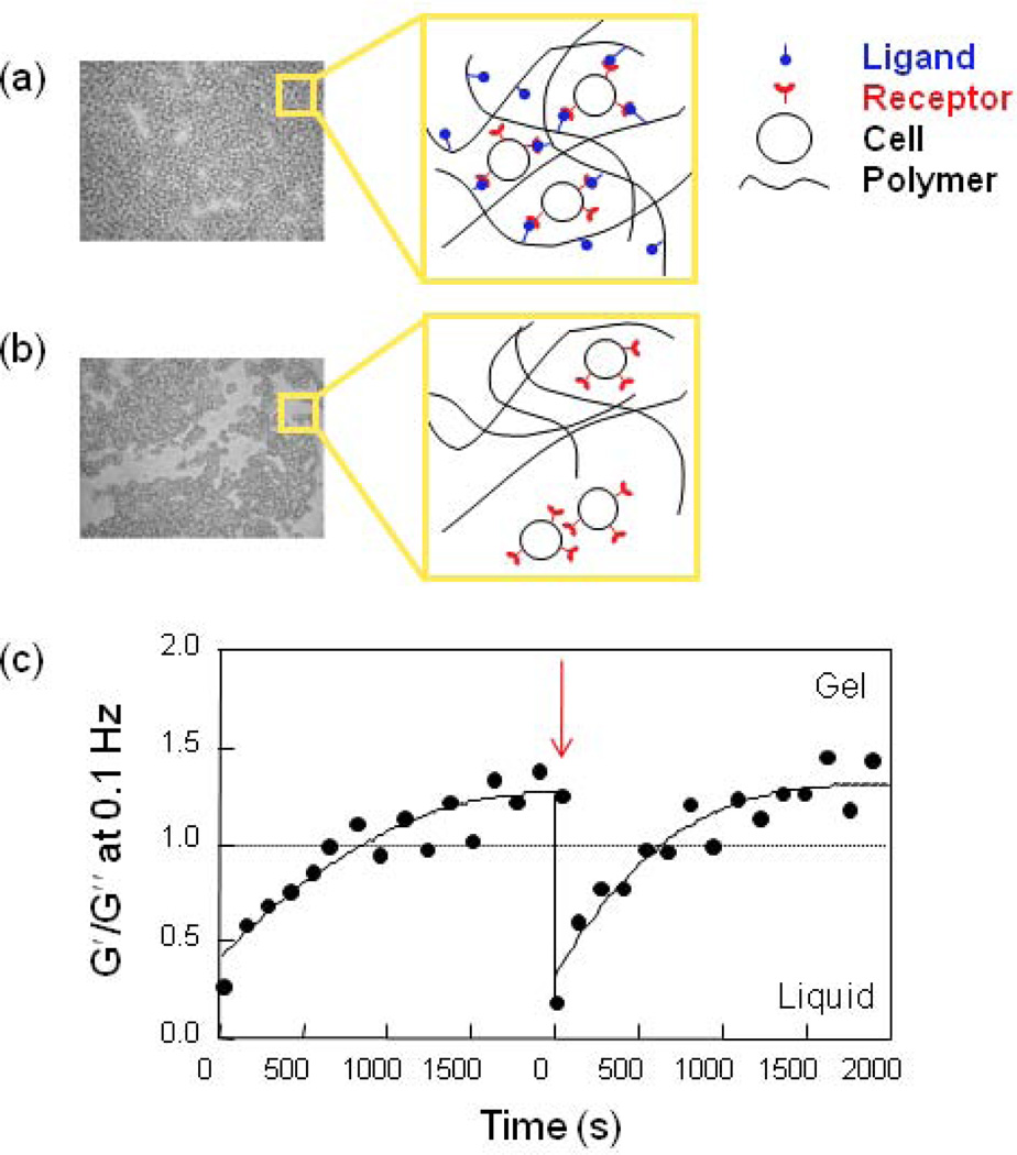
Photomicrographs and schematics of cell-cross-linked network structures when cells are mixed with (a) RGD-modified alginate and (b) non-modified alginate. (c) Shear-reversible gelation of cell-cross-linked RGD-alginate hydrogels. The arrow indicates disruption of the gel structure with intentional shear force. The cross-over point where G′ (storage shear modulus) = G″ (loss shear modulus) provides a measure of the gelation threshold (dotted line) [55]. Copyright 2003, WILEY-VCH Verlag GmbH & Co. KGaA, Weinheim, Germany.
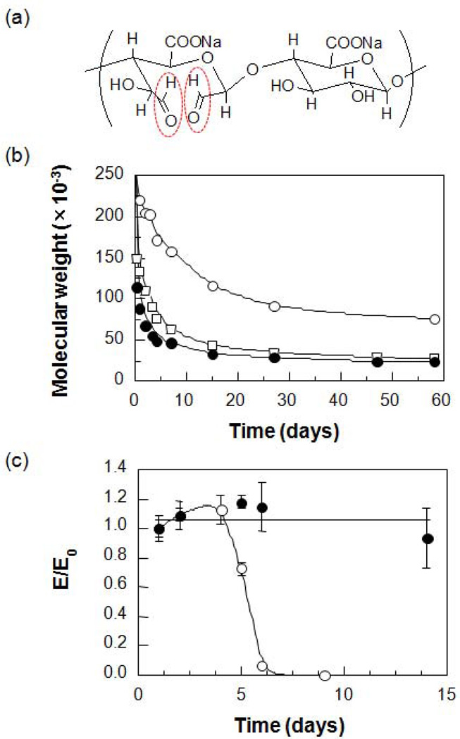
(a) Chemical structure of partially oxidized alginate (aldehyde groups generated after oxidation reaction highlighted in red) and (b) its degradation behavior at various pHs (○, pH 4.5; □, pH 7.4; ●, pH 9.2). (c) Changes in the compressive modulus of hydrogels prepared from alginate (●) and partially oxidized alginate (○) over time (pH 7.2, 37°C) [58]. Copyright 2001, John Wiley & Sons, New York, USA.
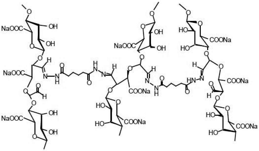
Chemical structure of poly(aldehyde guluronate) gels cross-linked with adipic acid dihydrazide [59]. Copyright 2000, American Chemical Society, Washington DC, USA.
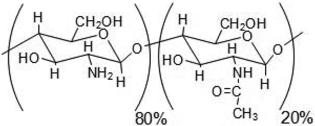
Chemical structure of chitosan.
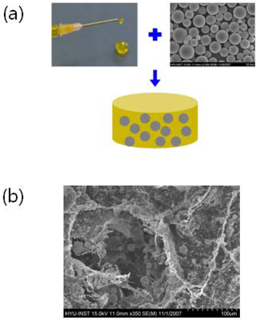
(a) A microsphere/hydrogel combination system can be prepared by ionic cross-linking of a suspension of poly(d,l-lactide-co-glycolide) (PLGA) microspheres containing protein drugs in an alginate solution. (b) Scanning electron microscopic image clearly shows even dispersion of PLGA microspheres in an alginate hydrogel [81]. Copyright 2009, WILEY-VCH Verlag GmbH & Co. KGaA, Weinheim, Germany.
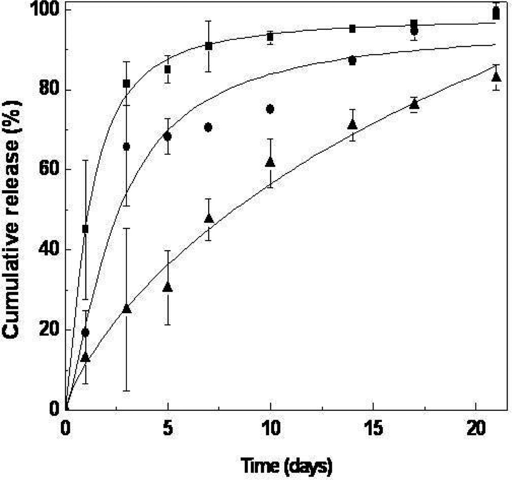
In vitro TAT-HSP27 release from microsphere/hydrogel combination delivery systems prepared at different mixing ratios (■, 0; ●, 1.0; ▲, 1.5; PLGA/alginate = w/w) [82]. Copyright 2009, Elsevier Science Ltd., Oxford, UK.
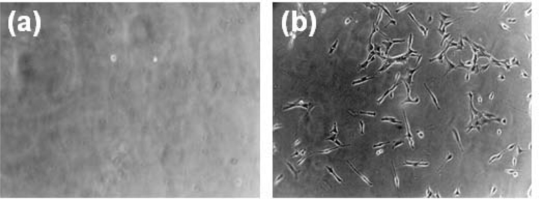
Optical microscope images of C2C12 myoblasts adhered to the surface of (a) non-peptide-modified alginate gels and (b) RGD-modified alginate gels. No cells attach and spread on the unmodified gels, while large numbers of well spread cells are found on the RGD-modified alginate. Images were taken after 24 hr culture at 100× magnification [98]. Copyright 1999, Elsevier Science Ltd., Oxford, UK.
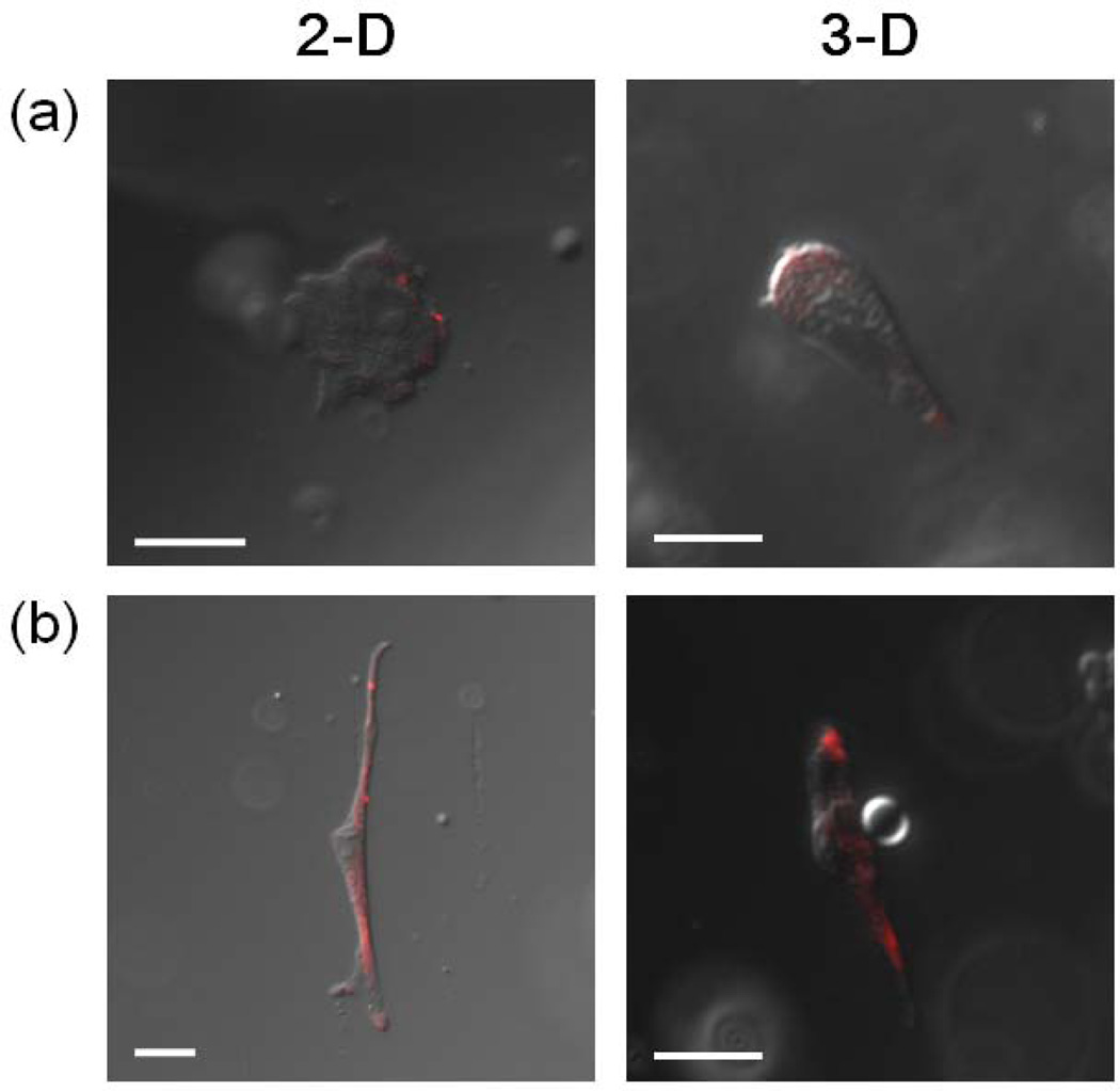
Confocal microscopic images of primary human fibroblasts cultured on alginate gels (2-D) modified with either (a) RGDSP or (b) G12RGDSP, and cells encapsulated within the same two types of gels (3-D). Images were taken after cells were treated with anti-vinculin antibody, followed by visualization with rhodamine-conjugated donkey anti-mouse IgG (scale bar, 20 µm) [99]. Cells cultured with G12RGDSP-alginate gels, both in 2-D and 3-D, clearly display focal contact formation, which is a sign of strong cell adhesion and regulates cell spreading and migration, as demonstrated by positive immunostaining for vinculin. Copyright 2010, Elsevier Science Ltd., Oxford, UK.
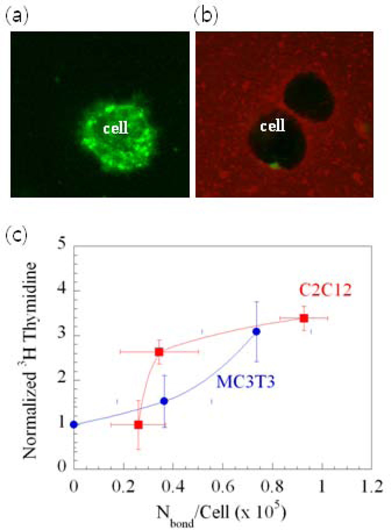
Directly visualizing and quantifying cell-gel adhesions, and their relation to cell phenotype. (a) A strong green emission of fluorescein in the cell membrane within unmodified gels was observed. (b) The color intensity of fluorescein in the cell membrane was greatly decreased and the color intensity of rhodamine at the interface between cells and gels was increased when cells were encapsulated in alginate gels presenting rhodamine-G4RGDASSKY, due to fluorescence resonance energy transfer (FRET). (c) The relationship between the amount of [3H]thymidine uptake by cells (indication of cell multiplication) and Nbond/cell for two cell types: muscle cells (C2C12) and bone cells (MC3T3-E1). The number of cell receptor–ligand bonds (Nbond) was determined using the FRET measurements [113]. Copyright 2006, National Academy of Sciences, Washington DC, USA.
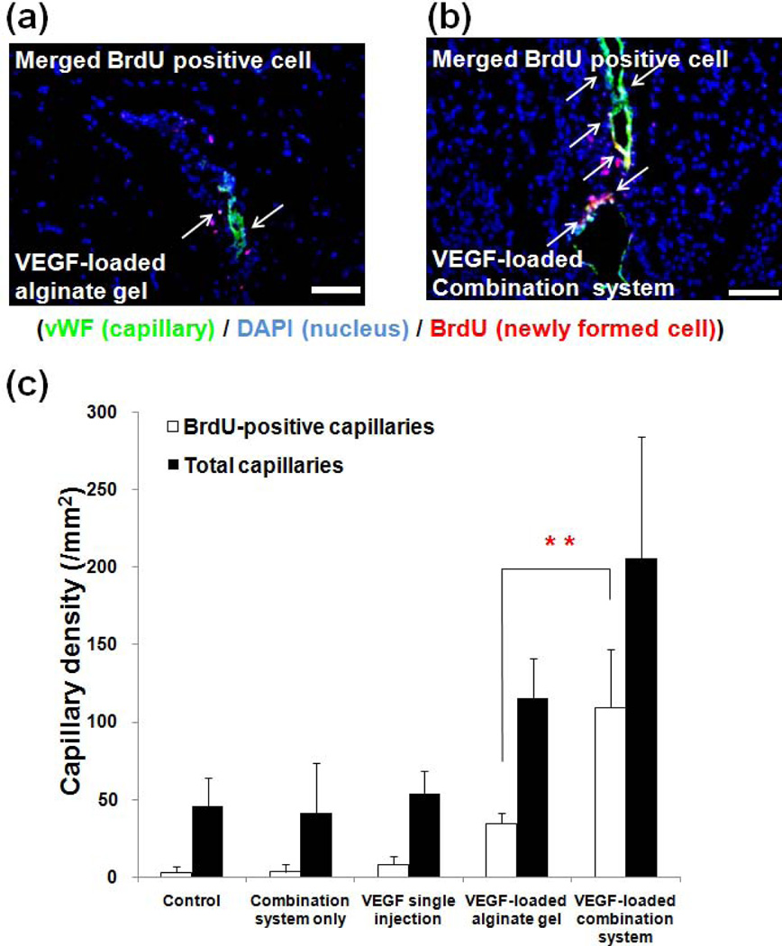
Tissues from mouse ischemic hindlimbs stained with anti-von Willebrand factor (vWF) (indicator of endothelial cells lining blood vessels) 4 weeks post-treatment with either (a) VEGF-loaded alginate hydrogel or (b) VEGF-loaded microsphere/hydrogel combination system. Blue, green and red colors represent DAPI (cell nuclei), vWF, and BrdU (indicating cells actively multiplying), respectively. Arrows indicate newly formed capillaries (scale bar, 20µm). (c) Quantification of the capillary density in tissue [121]. Copyright 2010, Springer, New York, USA.
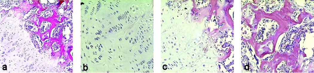
(a) Growth-plate-like structures formed by co-transplantation of chondrocytes and osteoblasts, similar to that observed in developing long bones (×100). Magnified images of the (b) cartilage, (c) transition, and (d) bone and marrow space regions (200× magnification) [130]. Copyright 2002, National Academy of Sciences, Washington DC, USA.
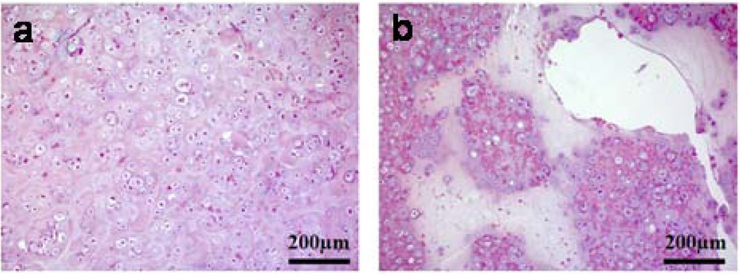
Images of H&E-stained tissues after 6 weeks following transplantation of primary chondrocytes into mice using either (a) cell cross-linked RGD-alginate gels or (b) media only [145]. Chondrocytes in lacunae (typically found in natural cartilage) were clearly observed for cell cross-linked RGD-alginate gels. Copyright 2009, WILEY-VCH Verlag GmbH & Co. KGaA, Weinheim, Germany.
References
-
- Ratner BD, Bryant SJ. Biomaterials: where we have been and where we are going? Ann Rev Biomed Eng. 2004;6:41–75. - PubMed
-
- Williams DF. On the nature of biomaterials. Biomaterials. 2009;30:5897–5909. - PubMed
-
- Gombotz WR, Wee SF. Protein release from alginate matrices. Adv Drug Delivery Rev. 1998;31:267–285. - PubMed
-
- Langer R, Vacanti JP. Tissue engineering. Science. 1993;260:920–926. - PubMed
Grants and funding
LinkOut - more resources
Full Text Sources
Other Literature Sources