. 2011 Nov;7(11):e1002368.
doi: 10.1371/journal.pgen.1002368. Epub 2011 Nov 17.

PBX1 genomic pioneer function drives ERα signaling underlying progression in breast cancer

Affiliations

PBX1 genomic pioneer function drives ERα signaling underlying progression in breast cancer

Luca Magnani et al. PLoS Genet. 2011 Nov.

Abstract

Altered transcriptional programs are a hallmark of diseases, yet how these are established is still ill-defined. PBX1 is a TALE homeodomain protein involved in the development of different types of cancers. The estrogen receptor alpha (ERα) is central to the development of two-thirds of all breast cancers. Here we demonstrate that PBX1 acts as a pioneer factor and is essential for the ERα-mediated transcriptional response driving aggressive tumors in breast cancer. Indeed, PBX1 expression correlates with ERα in primary breast tumors, and breast cancer cells depleted of PBX1 no longer proliferate following estrogen stimulation. Profiling PBX1 recruitment and chromatin accessibility across the genome of breast cancer cells through ChIP-seq and FAIRE-seq reveals that PBX1 is loaded and promotes chromatin openness at specific genomic locations through its capacity to read specific epigenetic signatures. Accordingly, PBX1 guides ERα recruitment to a specific subset of sites. Expression profiling studies demonstrate that PBX1 controls over 70% of the estrogen response. More importantly, the PBX1-dependent transcriptional program is associated with poor-outcome in breast cancer patients. Correspondingly, PBX1 expression alone can discriminate a priori the outcome in ERα-positive breast cancer patients. These features are markedly different from the previously characterized ERα-associated pioneer factor FoxA1. Indeed, PBX1 is the only pioneer factor identified to date that discriminates outcome such as metastasis in ERα-positive breast cancer patients. Together our results reveal that PBX1 is a novel pioneer factor defining aggressive ERα-positive breast tumors, as it guides ERα genomic activity to unique genomic regions promoting a transcriptional program favorable to breast cancer progression.


Conflict of interest statement

Patent pending for the use of PBX1 as a biomarker in breast cancer.

Figures
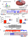
Figure 1
Figure 1. PBX1 correlates with ERα.
(A) Motif/sequence logo representation of the PBX1 matrix (Transfac: M01017). (B) The proportion of ERα binding sites harboring the PBX1 matrix (Transfac: M01017) is presented taking into account the overlap of ERα binding sites with FoxA1 binding sites. Percentages are calculated based on the 5782 ERα binding sites. (C) Co-expression of PBX1 and ERα mRNA transcripts is demonstrated across 47 distinct breast cancer cell lines separated based on their ERα-histological status. The p value revealing significant correlation between ERα-histological status and mRNA expression for ERα and PBX1 is presented. (D) Both mRNA (left panel) and protein (derived from immuno-fluorescence or western blot, right and bottom panel respectively) levels for PBX1 correlate with ERα expression status when assessed in ERα-positive (MCF7) and ERα–negative (MDA-MB231) breast cancer cells (average from three independent probes against PBX1 is presented for the mRNA expression analysis provided by bioGPS.org). (E) PBX1 and ERα are co-expressed in primary breast tumors. Expression profiles from primary breast tumors reveals that PBX1 mRNA levels are correlated with ERα-histological status and ERα mRNA expression in primary breast tumors (meta-analysis conducted using Oncomine). (*<0.05, **<0.01***, <0.001 p value).
Figure 2
Figure 2. PBX1 is required for the estrogen response in MCF7 cells.
(A) PBX1 depletion via siRNA effectively reduces its mRNA and (B) protein levels. (C) MCF7 breast cancer cells depleted of PBX1 fail to proliferate in response to estrogen/17β-estradiol (E2) stimulation compared to control treatment (O). (D) PBX1 silencing does not alter ERα or FoxA1 mRNA (histogram) or protein levels (Western Blot, WB). (*<0.05, **<0.01***, <0.001 p value).
Figure 3
Figure 3. PBX1 marks functional ERα bindings.
(A) Confocal immunofluorescence analysis in MCF7 cells cultured in absence of estrogen/17β-estradiol (E2) reveals that PBX1 is localized in the nucleus of MCF7 breast cancer cells and partially overlap with the pioneer factor FoxA1. (B) Venn diagram of PBX1 (Full media), ERα (after estrogen stimulation) and FoxA1 (full media) cistromes reveal their significant overlap on the chromatin. (C) A comparison between E2 responsive genes (all or PBX1-dependent) and the unique versus shared ERα, FoxA1 and PBX1 binding sites defined in Figure 3B was performed by normalizing the number of responsive genes with at least one unique or shared binding site a given factor within ±20 kb of their transcription start site (TSS) to the number of unresponsive genes with at least one binding sites from the same type of site within ±20 kb of their TSS. The values for all E2 responsive genes (blue line) and PBX1-dependent E2 responsive genes (red line) were plotted in a radar format (1< dark grey area, 1–2 light grey area, >2 white area, ticks are 0.5 increments). (*<0.01, **<0.001***, <0.00001 p value). (D) RT-qPCR against E2 target genes associated with PBX1-FoxA1-ERα, PBX1-ERα or FoxA1-ERα binding sites based on Figure 3C was performed in MCF7 breast cancer cells depleted of PBX1 (siPBX1) or Foxa1 (siFoxA1). A control siRNA (siCTRL) was used for comparison.
Figure 4
Figure 4. PBX1 is located in the nucleus and mediates ERα genomics activity.
(A) PBX1 occupies ERα genomic targets prior to its recruitment following estrogen/17β-estradiol (E2) stimulation compared to control treated cells (O). Similarly, PBX1 remains bound to the chromatin after E2 treatment in MCF7 breast cancer cells as determined by ChIP-qPCR. (B) ChIP-reChIP assays reveal that PBX1 and ERα co-occupy the same genomic regions upon E2 stimulation. In addition to a negative control site, matched IgG were used as a negative control in the reChIP assay. (C) PBX1 silencing (siPBX1) abrogates ERα recruitment at regulatory elements in MCF7 breast cancer cells compared to control (siCTRL). Values are calculated as a ratio between untreated and E2 treated relative fold enrichment defined by ChIP-qPCR. (D) ChIP-qPCR against ERα at PBX1-independent sites demonstrates that ERα recruitment is not disrupted at these sites upon PBX1 silencing. Values are calculated as a ratio between untreated and E2 treated relative fold enrichment.
Figure 5
Figure 5. PBX1 is an independent pioneer factor required for chromatin openness whose binding is favored by H3K4me2.
(A) Genome wide FAIRE profiles (FAIRE-seq) from MCF7 breast cancer cells maintained in estrogen-free media demonstrate that PBX1 alone or in combination with FoxA1 correlates with open chromatin. (B) Depletion of PBX1 (siPBX1) in MCF7 breast cancer cells maintained in estrogen-free media significantly reduces chromatin openness at PBX1 binding sites compared to control siRNA transfected cells (siCTRL) as measured by FAIRE-qPCR. (C) FoxA1 silencing (siFoxA1) does not alter PBX1 binding to the chromatin compared to control (siCTRL) in MCF7 breast cancer cells maintained in estrogen-free media. (D) PBX1 silencing in MCF7 breast cancer cells maintained in estrogen-free media does not affect FoxA1 binding to the chromatin compared to control. (E) Venn diagram of PBX1 and FoxA1 cistromes defined in full-media as well as H3K4me2 epigenome defined in MCF7 breast cancer cells maintained in estrogen-free media reveals their overlap. (F) Over-expression of the H3K4me2 demethylases KDM1 in MCF7 breast cancer cells maintained in estrogen-free media results in a significant reduction of PBX1 binding to the chromatin compared to the empty vector control (CTRL). (*<0.05, **<0.01***, <0.001 p value).
Figure 6
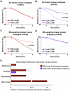
Figure 6. PBX1 is a novel prognostic marker for ERα positive breast cancers.
(A–B) PBX1 and FoxA1 prognostic value against metastasis-free survival were investigated against all breast cancer subtypes through Kaplan-Meier curves derived from a meta-analysis of independent expression profile studies from primary breast tumors available through Oncomine. (C–D) The same analysis was repeated while focusing only on the ERα-positive patients. Statistical difference in outcomes between patients with high (top 10% expressing patients) versus low (bottom 10% expressing patients) mRNA level was performed using Fisher exact test. (E) The number of expression signatures associated with poor-outcome defined in primary breast tumors in independent expression profile studies that are significantly associated with PBX1-dependent or FoxA1-dependent estrogen/17β-estradiol (E2) gene signatures is presented (p<0.01, O.R. >2). Results were generated using Oncomine concepts map analysis.
Figure 7
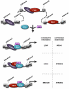
Figure 7. Schematic representation of PBX1 activity in breast cancer.
Schematic model depicting the relationship between PBX1, FoxA1 and ERα in breast cancer cells stimulated or not by estrogen/17β-estradiol (E2). Both FoxA1 and PBX1 are bound to the chromatin harboring the H3K4me2 epigenetic signature. They both act independently of each other to open chromatin making specific genomic regions accessible to transcription factors. Stimulation by E2 does not affect their chromatin occupancy but allows ERα recruitment. Importantly, sites of ERα recruitment bound by PBX1, shared or not with FoxA1, are associated with a significant proportion of estrogen responsive genes accounting for a strong estrogen response.

Comment in

  • Breast cancer: Staking a claim.
    McCarthy N. McCarthy N. Nat Rev Cancer. 2011 Dec 15;12(1):4. doi: 10.1038/nrc3192. Nat Rev Cancer. 2011. PMID: 22169975 No abstract available.

References

    1. Barrero MJ, Boue S, Izpisua Belmonte JC. Epigenetic mechanisms that regulate cell identity. Cell Stem Cell. 2010;7:565–570. - PubMed
    1. Meissner A. Epigenetic modifications in pluripotent and differentiated cells. Nature Biotechnology. 2010;28:1079–1088. - PubMed
    1. Bernstein BE, Mikkelsen TS, Xie X, Kamal M, Huebert DJ, et al. A bivalent chromatin structure marks key developmental genes in embryonic stem cells. Cell. 2006;125:315–326. - PubMed
    1. Meissner A, Mikkelsen TS, Gu H, Wernig M, Hanna J, et al. Genome-scale DNA methylation maps of pluripotent and differentiated cells. Nature. 2008;454:766–770. - PMC - PubMed
    1. Mikkelsen TS, Hanna J, Zhang X, Ku M, Wernig M, et al. Dissecting direct reprogramming through integrative genomic analysis. Nature. 2008;454:49–55. - PMC - PubMed

Publication types

MeSH terms

Substances