

Macular edema formation and deterioration of retinal function after intravitreal bevacizumab injection for proliferative diabetic retinopathy
- PMID: 22125532
- PMCID: PMC3220908
- DOI: 10.1159/000332830
Macular edema formation and deterioration of retinal function after intravitreal bevacizumab injection for proliferative diabetic retinopathy
Abstract
Purpose: To report a case of proliferative diabetic retinopathy (PDR) showing transient macular edema (ME) and deteriorated retinal function after intravitreal bevacizumab injection (IVB).
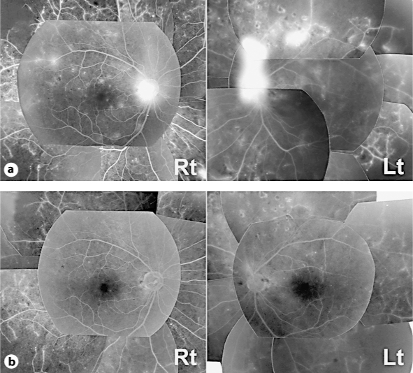
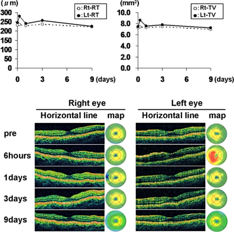
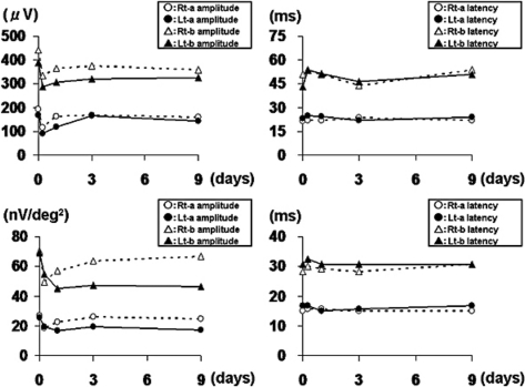
Methods and results: A 53-year-old man received IVB (1.25 mg/0.05 ml) in both eyes for the treatment of PDR. There was no treatment-related complication. However, he complained of photopsia in both eyes 6 h after the injection. Slit-lamp examination revealed mild cellular infiltrations (1+) in the anterior chamber in both eyes. Optical coherence tomography showed ME formation in the left eye. Both full-field and multifocal electroretinography (ERG) revealed the deterioration of all parameters in both eyes compared with pretreatment. The inflammation in the anterior segment and ME disappeared 1 day after the injection. ERG parameters were improved 9 days after the injection, except for the N1 and P1 amplitude of multifocal ERG in the left eye.
Conclusion: We propose that patients who undergo IVB should be carefully informed and followed up for possible complications including temporal ME formation and retinal function deterioration.
Keywords: Anti-vascular endothelial growth factor; Avastin; Bevacizumab; Diabetic retinopathy; Full-field electroretinography; Macular edema; Multifocal electroretinography; Optical coherence tomography.
Figures
Similar articles
-
Prospective study of intravitreal triamcinolone acetonide versus bevacizumab for macular edema secondary to central retinal vein occlusion.Retina. 2011 May;31(5):838-45. doi: 10.1097/IAE.0b013e3181f4420d. Retina. 2011. PMID: 21293319 Clinical Trial.
-
Electrophysiological and structural assessment of the central retina following intravitreal injection of bevacizumab for treatment of macular edema.Doc Ophthalmol. 2008 Mar;116(2):129-35. doi: 10.1007/s10633-007-9090-9. Epub 2007 Oct 25. Doc Ophthalmol. 2008. PMID: 17960440
-
Intravitreal bevacizumab (Avastin) therapy for persistent diffuse diabetic macular edema.Retina. 2006 Nov-Dec;26(9):999-1005. doi: 10.1097/01.iae.0000247165.38655.bf. Retina. 2006. PMID: 17151486 Clinical Trial.
-
Intravitreal bevacizumab (avastin) injection alone or combined with triamcinolone versus macular photocoagulation as primary treatment of diabetic macular edema.Retina. 2007 Nov-Dec;27(9):1187-95. doi: 10.1097/IAE.0b013e31815ec261. Retina. 2007. PMID: 18046223 Clinical Trial.
-
Extrafoveal changes following intravitreal bevacizumab injections for macular edema secondary to branch retinal vein occlusion: an mfERG and OCT study.Doc Ophthalmol. 2013 Apr;126(2):137-48. doi: 10.1007/s10633-012-9367-5. Epub 2012 Dec 20. Doc Ophthalmol. 2013. PMID: 23255086
References
-
- Arevalo JF, Wu L, Sanchez JG, et al. Intravitreal bevacizumab (avastin) for proliferative diabetic retinopathy: 6-months follow-up. Eye. 2009;23:117–123. - PubMed
-
- Ishikawa K, Honda S, Tsukahara Y, Negi A. Preferable use of intravitreal bevacizumab as a pretreatment of vitrectomy for severe proliferative diabetic retinopathy. Eye. 2009;23:108–111. - PubMed
-
- Modarres M, Nazari H, Falavarjani KG, Naseripour M, Hashemi M, Parvaresh MM. Intravitreal injection of bevacizumab before vitrectomy for proliferative diabetic retinopathy. Eur J Ophthalmol. 2009;19:848–852. - PubMed
-
- Arevalo JF, Maia M, Flynn HW, Jr, Saravia M, Avery RL, Wu L, Eid Farah M, DJ Pieramici, MH Berrocal, JG Sanchez. Tractional retinal detachment following intravitreal bevacizumab (Avastin) in patients with severe proliferative diabetic retinopathy. Br J Ophthalmol. 2008;92:213–216. - PubMed
-
- Jonas JB, Schmidbauer M, Rensch F. Progression of tractional retinal detachment following intravitreal bevacizumab. Acta Ophthalmol. 2009;87:571–572. - PubMed
Publication types
LinkOut - more resources
Full Text Sources
