. 2011;6(11):e26674.
doi: 10.1371/journal.pone.0026674. Epub 2011 Nov 17.

Suppressor of cytokine signalling-6 promotes neurite outgrowth via JAK2/STAT5-mediated signalling pathway, involving negative feedback inhibition

Affiliations

Suppressor of cytokine signalling-6 promotes neurite outgrowth via JAK2/STAT5-mediated signalling pathway, involving negative feedback inhibition

Sakshi Gupta et al. PLoS One. 2011.

Abstract

Background: Suppressors of cytokine signalling (SOCS) protein family are key regulators of cellular responses to cytokines and play an important role in the nervous system. The SOCS6 protein, a less extensively studied SOCS family member, has been shown to induce insulin resistance in the retina and promote survival of the retinal neurons. But no reports are available about the role of SOCS6 in neuritogenesis. In this study, we examined the role of SOCS6 in neurite outgrowth and neuronal cell signalling.

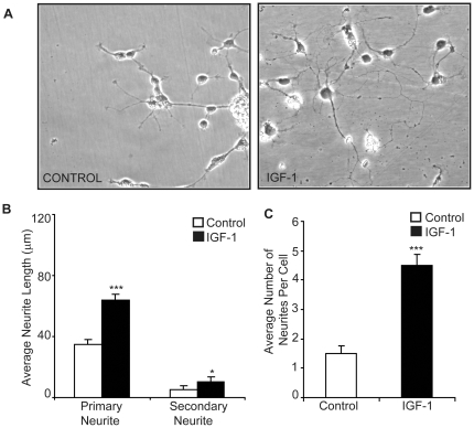
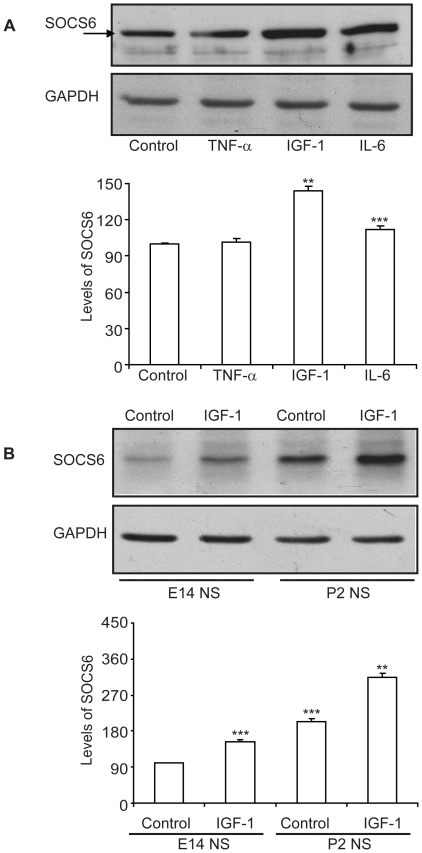
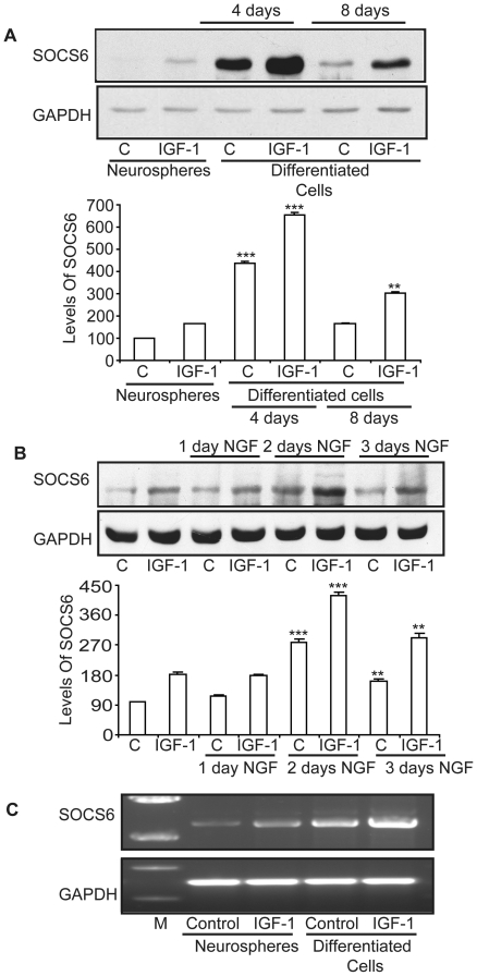
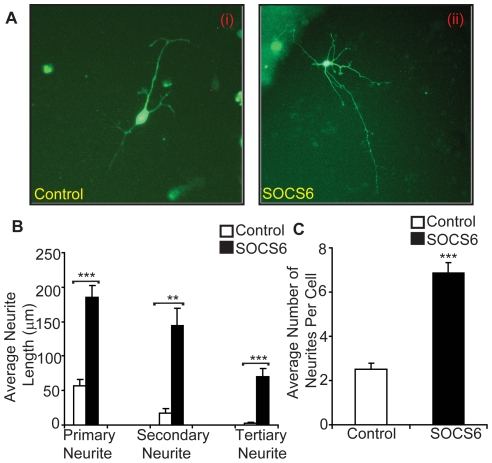
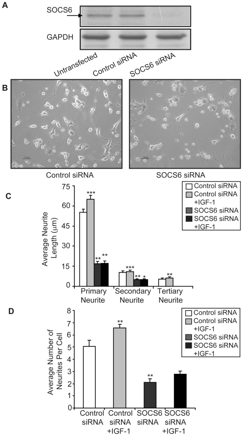
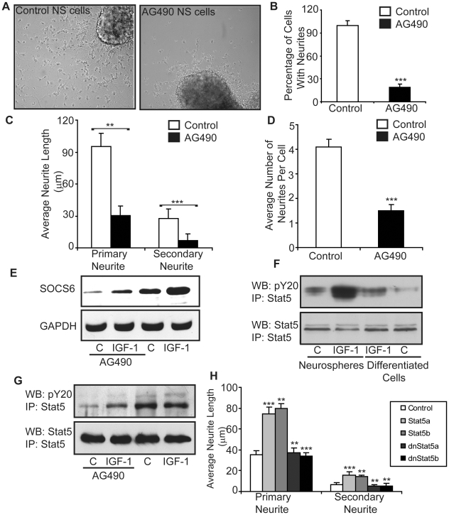
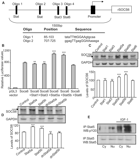
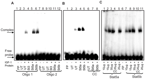
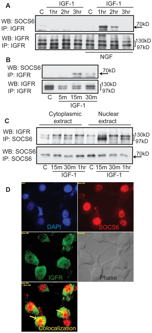
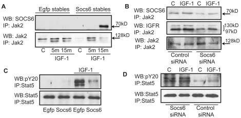
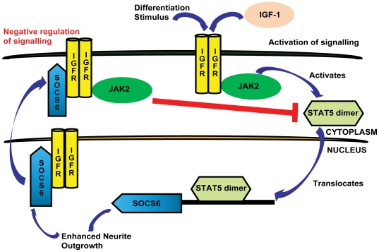
Methodology/principal findings: The effect of SOCS6 in neural stem cells differentiation was studied in neural stem cells and PC12 cell line. Highly elevated levels of SOCS6 were found upon neural cell differentiation both at the mRNA and protein level. Furthermore, SOCS6 over-expression lead to increase in neurite outgrowth and degree of branching, whereas SOCS6 knockdown with specific siRNAs, lead to a significant decrease in neurite initiation and extension. Insulin-like growth factor-1 (IGF-1) stimulation which enhanced neurite outgrowth of neural cells resulted in further enhancement of SOCS6 expression. Jak/Stat (Janus Kinase/Signal Transducer And Activator Of Transcription) pathway was found to be involved in the SOCS6 mediated neurite outgrowth. Bioinformatics study revealed presence of putative Stat binding sites in the SOCS6 promoter region. Transcription factors Stat5a and Stat5b were involved in SOCS6 gene upregulation leading to neuronal differentiation. Following differentiation, SOCS6 was found to form a ternary complex with IGFR (Insulin Like Growth Factor-1 Receptor) and JAK2 which acted in a negative feedback loop to inhibit pStat5 activation.

Conclusion/significance: The current paradigm for the first time states that SOCS6, a SOCS family member, plays an important role in the process of neuronal differentiation. These findings define a novel molecular mechanism for Jak2/Stat5 mediated SOCS6 signalling.


Conflict of interest statement

Competing Interests: The authors have declared that no competing interests exist.

Figures

Figure 1
Figure 1. IGF-1 enhances neurite-outgrowth.
(A) E14 neurospheres were allowed to differentiate in the presence or absence of IGF-1. (B, C) Analysis of average of primary and secondary neurite length and average number of neurites per cell in NSCs differentiated in presence and absence of IGF-1. Neurite length was measured in randomly chosen cells (at least 8–10 different fields and approx 5 cells per field) by tracing individual neurites (as described in experimental procedures) and results are expressed as (B) average of total primary and secondary neurite lengths or (C) average number of neurites per cell. Statistical significance of the difference was determined using ANOVA. The result shows the mean ± S.E. of n = 3 combined experiments (***p<0.001, *p<0.05).
Figure 2
Figure 2. IGF-1 stimulation enhances SOCS6 levels.
(A) Primary neurospheres were generated from cortex of E14 rat embryos and stimulated with TNF-α (100 pg/ml), IL-6 (20 ng/ml) or IGF-1 (20 ng/ml) for three hours. The cell lysate was immunoblotted with anti-SOCS6 antibody. The same membrane was stripped and reprobed with anti-GAPDH antibody for protein loading control. (B) Primary neurospheres were generated from cortex of E14 and sub ventricular zone of pups at postnatal day two (P2) and stimulated with/without IGF-1 for 3 hours. The cell lysate was immunoblotted with anti-SOCS6 antibody. The same membrane was stripped and reprobed with anti-GAPDH antibody for protein loading control. The densitometry data shown was normalized with the untreated control (taken as 100%). The result shows the mean ± S.E. of n = 3 combined experiments (***p<0.001, **p<0.01).
Figure 3
Figure 3. Temporal increase in SOCS6 levels following differentiation.
(A) Western-blot analysis of SOCS6 expression in E14 neurospheres and neurospheres upon differentiation at day 4 and day 8 with/without 3 hours of IGF-1 (20 ng/ml) stimulation. The same membrane was stripped and reprobed with anti-GAPDH antibody for protein loading control. (B). Western-blot analysis of SOCS6 expression in undifferentiated PC12 cells and PC12 cells differentiated with NGF (50 ng/ml) for 1, 2 or 3 days and stimulated with/without IGF-1 for 3 hours. The same membrane was stripped and reprobed with anti-GAPDH antibody for protein loading control. (C) E14 neurospheres or neurospheres upon 4 days of differentiation were stimulated with/without IGF-1. Total RNA was isolated and RT-PCR was performed using SOCS6 specific primers and GAPDH primers on the same sample. (M = Marker; C = Control). The result shows the mean ± S.E. of n = 3 combined experiments (***p<0.001, **p<0.01). The densitometry shown below was normalized with the untreated control (taken as 100%).
Figure 4
Figure 4. SOCS6 enhances neurite-outgrowth.
(A) Following withdrawal of growth factors, E14 neurospheres were transiently transfected with control (EGFP vector alone) (i) or SOCS6 (SOCS6-EGFP) (ii) and observed under fluorescent microscope. (B, C) Neurite length was measured in randomly chosen cells (at least 6–8 different fields and approx 5 cells per field) by tracing individual neurites (as described in experimental procedures) and results are expressed as (B) average of total primary, secondary and tertiary neurite lengths or (C) average number of neurites per cell. Statistical significance of the difference was determined using ANOVA. The result shows the mean ± S.E. of n = 3 combined experiments (***p<0.001, **p<0.01).
Figure 5
Figure 5. siRNA-mediated silencing of SOCS6 expression inhibits neurite-outgrowth.
(A) 3 µg of siRNA was transfected into PC12 cells and after 6 days of transfection, the cells were screened for SOCS6 knockdown by Western-blot analysis, using anti-SOCS6 antibody. Control-siRNA (green non-targeting) was used as a negative control. The same membrane was stripped and reprobed with anti-GAPDH antibody for protein loading control. (B) Morphology of SOCS6-siRNA transfected PC12 cells as compared to the control transfected cells after 6 days of transfection with NGF treatment under light microscope. (C and D) Neurite length was measured in randomly chosen cells (at least 8–10 different fields and approx 5 cells per field) transfected with either control or SOCS6 siRNA with or without IGF-1. Neurite lengths were measured by tracing individual neurites (as described in experimental procedures) and results are expressed as (C) average of total primary, secondary and tertiary neurite lengths or (D) average number of neurites per cell. Statistical significance of the difference was determined using ANOVA. The result shows the mean ± S.E. of n = 3 combined experiments (***p<0.001, **p<0.01, *p<0.05).
Figure 6
Figure 6. Jak/Stat pathway is involved in neural stem cell differentiation.
(A) E14 Neurospheres were allowed to differentiate in the absence or presence of 50 µM AG490 (Jak2/Stat3 inhibitor) for 4 days and the cells were observed under light microscope. (B) Number of cells with neurites per field was counted. An average of 13 fields was taken. The untreated control was taken as 100%. (C and D) Neurite length was measured in randomly chosen cells by tracing individual neurites (as described in experimental procedures) and results are expressed as (C) average of total primary and secondary neurite lengths. The untreated control was taken as 100%. (D) Average number of neurites per cell. (E) Untreated or AG490 treated neurospheres were stimulated with/without IGF-1. The cell lysate was immunoblotted with anti-SOCS6 antibody. The membrane was then stripped and reprobed with anti-GAPDH antibody. (F) E14 Neurospheres and neurospheres upon differentiation were stimulated with/without IGF-1 for 10 minutes. Stat5 was immunoprecipitated from 300 µg protein extract and Western-blotted using anti-phosphotyrosine antibody (pY20). The membrane was then stripped and reprobed with anti-Stat5 antibody. (G) Untreated or AG490 treated neurospheres were stimulated with/without IGF-1 for 10 minutes. Stat5 was immunoprecipitated from 300 µg protein extract and Western-blotted using anti-phosphotyrosine antibody (pY20). The membrane was then stripped and reprobed with anti-Stat5 antibody. (H) PC12 cells, transfected with Stat5a- pcDNA3.1, Stat5b- pcDNA3.1, dominant negative Stat5a- pcDNA3.1 and dominant negative Stat5b- pcDNA3.1 were allowed to differentiate in the presence of NGF and the cells were observed under light microscope. Neurite length was measured in randomly chosen cells by tracing individual neurites (as described in experimental procedures) and results are expressed as average of total primary and secondary neurite lengths. Statistical significance of the difference was determined using ANOVA. The result shows the mean ± S.E. of n = 3 combined experiments (***p<0.001, **p<0.01).
Figure 7
Figure 7. Stat5a and Stat5b upregulate SOCS6 expression.
(A) Top panel is a diagrammatic representation of the 1500 bp upstream region of rat SOCS6 promoter showing the 2 putative unknown STAT binding sites, one Stat3 and one Stat6 binding sites. Oligo 1 and Oligo 2 are the two oligomers used for the EMSA binding assays. The lower panel displays the sequence of the two oligos based on the Genomatix software predicting the putative STAT binding sites. (B) PC12 cells were co-transfected with 1500 bp-pGL3 SOCS6, β-galactosidase plasmid and specific Stat-pcDNA3.1 constructs (Stat1, Stat3, Stat5a, Stat5b, and Stat6). Subsequently a luciferase assay was performed as described in experimental procedures. Each assay was performed in triplicate. (C) PC12 cells were transfected with pcDNA3.1-Stat1, pcDNA3.1-Stat3, pcDNA3.1-Stat5a, pcDNA3.1-Stat5b and pcDNA3.1-Stat6. After 2 days, lysates were prepared and the cell lysate was immunoblotted with anti-SOCS6 antibody. The membrane was subsequently stripped and reprobed with anti-GAPDH antibody for protein loading control. (D) PC12 cells were transfected with pcDNA3.1-Stat5a, pcDNA3.1-Stat5b, dominant negative pcDNA3.1-Stat5a and dominant negative pcDNA3.1-Stat5b. After 2 days, lysates were prepared and the cell lysate was immunoblotted with anti-SOCS6 antibody and subsequently stripped and reprobed with anti-GAPDH antibody for protein loading control. (E) PC12 cells were stimulated with/without IGF-1 and nuclear and cytoplasmic protein was extracted (as described in experimental procedures). Immunoprecipitation was performed with anti-Stat5 antibody and Western-blotted with phosphotyrosine (pY20) antibody. The membrane was stripped and reprobed with anti-Stat5 antibody for loading control. (Cy = Cytoplasmic; Nu = Nuclear). The densitometry shown below was normalized with the untreated control (taken as 100%). The result shows the mean ± S.E. of n = 3 combined experiments (***p<0.001).
Figure 8
Figure 8. Stat5a and Stat5b are the transcription factors for SOCS6 promoter.
(A) EMSA with Oligo 1 and Oligo 2 was performed using nuclear extracts from untransfected and PC12 cells transfected with Stat3-EGFP, Stat5a-EGFP and Stat5b-EGFP and stimulated with IGF-1 for 1 hour. Oligo 1 was used in lanes 1–6 and Oligo 2 in lanes 7–12. Lane 1, without cell extracts (free probe); lane 2, negative control (untransfected and without IGF-1 stimulation); lane 3, control (untransfected and with IGF-1 stimulation); lane 4, Stat3-EGFP; lane 5, Stat5a-EGFP; lane 6, Stat5b-EGFP; lane 7, without cell extracts (free probe); lane 8, negative control (untransfected and without IGF-1 stimulation); lane 9, control (untransfected and with IGF-1 stimulation); lane 10, Stat3-EGFP; lane 11, Stat5a-EGFP; lane 12, Stat5b-EGFP. (B) EMSA with Oligo1 with cold competition assay. Specificity of binding was checked using excess of cold oligomer as a competitor. Lane 1, without cell extracts; lane 2, negative control (untransfected and without IGF-1 stimulation); lane 3, control (untransfected and with IGF-1 stimulation); lane 4, Stat5a-EGFP; lane 5, Stat5b-EGFP; lanes (6–8), competition assay with excess of cold Oligo 1; lane 6, control (untransfected and with IGF-1 stimulation); lane 7, Stat5a-EGFP; lane 8, Stat5b-EGFP. (C) EMSA with Olig 1. Specificity of binding was further checked by using a protein gradient. Lanes 1–5 were Stat5a transfected and lanes 7–11 were Stat5b transfected and IGF-1 stimulated cell extracts. Lane 1, 10 µg protein; lane 2, 15 µg protein; lane 3, 30 µg protein; lane 4, competition assay with cold probe (20 µg protein); lane 5, 20 µg protein; lane 6, without cell extracts; lane 7, 10 µg protein; lane 8, 15 µg protein; lane 9, 30 µg protein; lane 10, competition assay with cold probe (20 µg protein); lane 11, 20 µg protein. The positions of free probe and specific complex are indicated. All the experiments were independently repeated 3 times with similar results. Free probe = FP, untransfected = UT.
Figure 9
Figure 9. SOCS6 associates with IGFR upon IGF-1 stimulation.
(A) Undifferentiated or NGF differentiated PC12 cells were stimulated with/without IGF-1 for 1, 2 or 3 hours. Using 300 µg of cell lysate, IGFR was pulled down and Western-blotted with anti-SOCS6 antibody. The membrane was stripped and reprobed with anti-IGFR antibody for loading control. (B) SOCS6 stable PC12 cells were stimulated with/without IGF-1 for 5, 15, and 30 minutes. IGFR was immunoprecipitated and Western-blotted with anti-SOCS6 antibody. The membrane was stripped and reprobed with anti-IGFR antibody for loading control. (C) PC12 cells were differentiated for 2 days with NGF and then stimulated with/without IGF-1 for 15, 30 m or 1 hr (m = minutes and hr = hours) and nuclear and cytoplasmic protein was extracted (as described in experimental procedures). Using 300 µg of cell lysate, SOCS6 was pulled down and Western-blotted with anti-IGFR antibody. The membrane was stripped and reprobed with anti-SOCS6 antibody for loading control. (D) PC12 cells were allowed to differentiate for 2 days with NGF and stimulated with IGF-1 for 30 minutes. The cells were then fixed and permeabilized. After primary antibody treatment (anti-SOCS6, anti-IGFR and anti-SOCS6+anti-IGFR), SOCS6 was stained with Alexafluor 594 (red) and IGFR was stained with Alexafluor 488 (green). The cells were visualized under fluorescent microscope. DAPI staining shows the location of the nucleus.
Figure 10
Figure 10. Feedback inhibition of Jak2/Stat5 pathway occurs via formation of a Jak2-IGFR-SOCS6 complex.
(A) PC12 cells stably transfected with SOCS6-EGFP or EGFP vector alone were stimulated with IGF-1 for 0 m, 5 m and 15 m (m = minute). Jak2 was immunoprecipitated and Western-blotted with anti-SOCS6 (top panel) or anti-Jak2 (bottom panel) antibodies. (B) PC12 cells transiently transfected with control or SOCS6-siRNA was stimulated with IGF-1 for 15 minutes. Jak2 was immunoprecipitated and Western-blot was performed using anti-IGFR antibody. The membrane was subsequently stripped and reprobed with anti-SOCS6 and anti-Jak2 antibodies. (C) PC12 cells stably transfected with SOCS6-EGFP or EGFP vector alone were stimulated with IGF-1 for 10 minutes. Stat5 was immunoprecipitated and Western-blot was performed using pY20 antibody. The membrane was subsequently stripped and reprobed with anti-Stat5 antibody. (D) PC12 cells transiently transfected with control or SOCS6-siRNA was stimulated with IGF-1 for 10 minutes. Stat5 was immunoprecipitated and Western-blot was performed using pY20 antibody. The membrane was subsequently stripped and reprobed with anti-Stat5 antibody. All the experiments were independently repeated 3 times with similar results.
Figure 11
Figure 11. Schematic representation of SOCS6 mediated neural differentiation.
Upon IGF-1 stimulation of neural cells, Jak2 is activated which then activates Stat5. Phosphorylated Stat5 dimerizes, translocates to the nucleus and acts as a transcription factor for SOCS6 which in turn promotes differentiation. SOCS6 then binds to IGFR and forms a trio complex with Jak2 leading to the inhibition of further Jak2-Stat5 signalling.

Similar articles

Cited by

References

    1. da Silva JS, Dotti CG. Breaking the neuronal sphere: regulation of the actin cytoskeleton in neuritogenesis. Nat Rev Neurosci. 2002;3:694–704. - PubMed
    1. Goldshmit Y, Galea MP, Wise G, Bartlett PF, Turnley AM. Axonal regeneration and lack of astrocytic gliosis in EphA4-deficient mice. J Neurosci. 2004;24:10064–10073. - PMC - PubMed
    1. Goldshmit Y, Greenhalgh CJ, Turnley AM. Suppressor of cytokine signalling-2 and epidermal growth factor regulate neurite outgrowth of cortical neurons. Eur J Neurosci. 2004;20:2260–22660. - PubMed
    1. Goldshmit Y, Walters CE, Scott HJ, Greenhalgh CJ, Turnley AM. SOCS2 induces neurite outgrowth by regulation of epidermal growth factor receptor activation. J Biol Chem. 2004;279:16349–16355. - PubMed
    1. Krebs DL, Hilton DJ. SOCS: physiological suppressors of cytokine signalling. J Cell Sci. 2000;113:2813–2819. - PubMed

Publication types

MeSH terms