. 2011;6(11):e26898.
doi: 10.1371/journal.pone.0026898. Epub 2011 Nov 17.

Intestinal subepithelial myofibroblasts support in vitro and in vivo growth of human small intestinal epithelium

Affiliations

Intestinal subepithelial myofibroblasts support in vitro and in vivo growth of human small intestinal epithelium

Nicholas Lahar et al. PLoS One. 2011.

Abstract

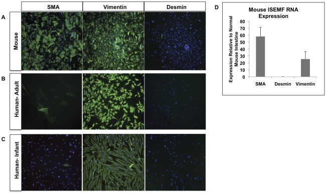
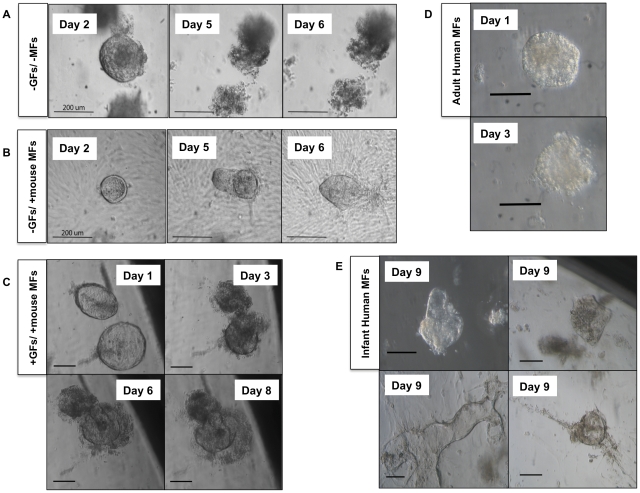
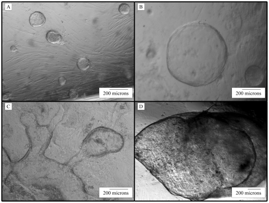
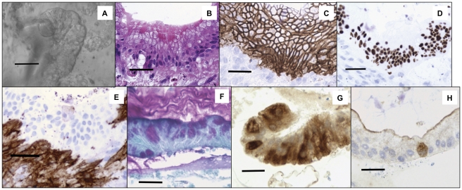
The intestinal crypt-niche interaction is thought to be essential to the function, maintenance, and proliferation of progenitor stem cells found at the bases of intestinal crypts. These stem cells are constantly renewing the intestinal epithelium by sending differentiated cells from the base of the crypts of Lieberkühn to the villus tips where they slough off into the intestinal lumen. The intestinal niche consists of various cell types, extracellular matrix, and growth factors and surrounds the intestinal progenitor cells. There have recently been advances in the understanding of the interactions that regulate the behavior of the intestinal epithelium and there is great interest in methods for isolating and expanding viable intestinal epithelium. However, there is no method to maintain primary human small intestinal epithelium in culture over a prolonged period of time. Similarly no method has been published that describes isolation and support of human intestinal epithelium in an in vivo model. We describe a technique to isolate and maintain human small intestinal epithelium in vitro from surgical specimens. We also describe a novel method to maintain human intestinal epithelium subcutaneously in a mouse model for a prolonged period of time. Our methods require various growth factors and the intimate interaction between intestinal sub-epithelial myofibroblasts (ISEMFs) and the intestinal epithelial cells to support the epithelial in vitro and in vivo growth. Absence of these myofibroblasts precluded successful maintenance of epithelial cell formation and proliferation beyond just a few days, even in the presence of supportive growth factors. We believe that the methods described here can be used to explore the molecular basis of human intestinal stem cell support, maintenance, and growth.


Conflict of interest statement

Competing Interests: The authors have declared that no competing interests exist.

Figures

Figure 1
Figure 1. Characterization of Mouse and Human Myofibroblasts.
(A) C57 BL/6 murine myofibroblasts plated in plastic culture dishes for four days. Using immunofluorescence, cells stained characteristically for intestinal myofibroblasts with positive SMA and vimentin staining and negative desmin staining. The blue pseudocolor is DAPI counterstaining for cell nuclei. (B) Myofibroblasts were isolated from adult human ileostomy surgical samples and plated for four days prior to immunofluorescence staining. Similar to murine myofibroblasts, SMA and vimentin stains were positive while the desmin stain was negative. (C) Myofibroblasts were isolated from a human infant ileostomy and plated for four days prior to immunofluorescence staining. Like the adult human sample, SMA was positive but faint. Vimentin stains were positive while the desmin stain was negative. (D) PCR results performed on the C57 BL/6 murine myofibroblasts consistent with the immunofluorescence results of positive SMA and vimentin staining and negative desmin staining.
Figure 2
Figure 2. Supportive Effect of Wnt, FGF Growth Factors and Myofibroblasts on Human Epithelial Growth.
(A) Human small intestinal crypts cultured without myofibroblasts (MFs) or growth factors (GFs) will live for approximately two days before dying off. (B) Human small intestinal crypts cultured in the presence of murine myofibroblast but without growth factors maintain their cystic shape indefinitely but without significant growth. (C) Human small intestinal crypts cultured with Wnt3a and FGF10 growth factors in the presence of mouse myofibroblasts began as simple cysts that fill and extruded their contents and became more complex in morphology over time. (D) Human epithelial clusters grown on adult human myofibroblasts in the presence of growth factors organize into simple cysts but cannot be maintained for longer than 3 days. (E) Infant human myofibroblasts are capable of supporting human intestinal epithelial growth into complex cystic structures through 9 days post-explantation. For A and B, scale bar is 200 µm. For C, D, and E, scale bar is 100 µm.
Figure 3
Figure 3. Long-Term Culture of Human Small Intestinal Enteroids Grown on Human Infant Myofibroblasts.
Micrographs of human small intestinal crypts that were cultured on human infant myofibroblasts for (A) 2 days, (B) 8 days, (C) 23 days, and (D) 56 days in culture. Small cysts get larger with time and sometimes fuse with each other to form larger structures.
Figure 4
Figure 4. In vitro Human Small Intestinal Enteroids Grown on Murine Myofibroblasts Demonstrate Characteristic Intestinal Epithelial Markers.
Human crypts cultured on murine myofibroblasts in the presence of Wnt3a and FGF10 were processed after 18 days in vitro. (A) Phase contrast microscopy of culture. (B) Hematoxylin and Eosin stain. Note cellular polarity with epithelial nuclei at the basal region and goblet cells at the apical region. (C) E-cadherin, an epithelial cell marker. (D) CDX-2, stains intestinal epithelium. Of note, while the E-cadherin staining is relatively even, the CDX-2 staining demonstrates uneven staining suggestive of alternating crypt-villus domains. (E) Smooth Muscle Actin, marker for myofibroblasts. (F) PAS, stains for goblet cells. Note the extruded mucinous material at the apical side of the epithelium culture. (G) Lysozyme, a Paneth cell marker. (H) Synaptophysin, marker for enteroendocrine cells. For all images, scale bars are 100 µm.
Figure 5
Figure 5. Long-term In vitro Human Small Intestinal Enteroids Grown on Murine Myofibroblasts.
Human small intestinal crypts cultured on murine myofibroblasts in the presence of Wnt3a and FGF10 were processed after 58 days in vitro. (A) Hematoxylin and Eosin stain. (B) CDX-2, stains intestinal epithelium. (C) E-cadherin, an epithelial cell marker. (D) α Smooth Muscle Actin, marker for myofibroblasts.
Figure 6
Figure 6. In vivo Human Small Intestinal Enteroids Can Be Maintained with Murine ISEMFs.
Human small intestinal crypts on murine myofibroblasts were grown in culture for 11 days in the presence of Wnt3a and FGF10, and then placed on a PGA felt scaffold and implanted subcutaneously into an immunocompromised NOD-SCID-IL2Rγ null mice. After 28 days, the implant was harvested and evaluated with intestinal epithelial markers. (A) H&E demonstrates at least three cell morphologies, and eosinophilic material in the cyst lumen. (B) E-cadherin and (C) CDX-2, are intestinal epithelial cell markers. Again note the variable staining intensity by the CDX-2 suggestive of crypt and villus domains (D) α Smooth Muscle Actin staining for myofibroblast adjacent to epithelial cells. (E) PAS staining for mucin and mucin producing goblet cells (F) Chromogranin A, marker for enteroendocrine cells. For all images, scale bar is 100 µm.
Figure 7
Figure 7. In vivo Human Small Intestinal Enteroids Can Be Maintained with Human Infant ISEMFs.
Human small intestinal crypts were grown on human myofibroblasts for 8 days, and then placed on a PGA felt scaffold and implanted subcutaneously into an immunocompromised NOD-SCID-IL2Rγ null mice. After 28 days, the implant was harvested and evaluated with intestinal epithelial markers. (A) H&E staining showing epithelial organization. (B) E-cadherin and (C) CDX-2 are intestinal epithelial cell markers. (D) α Smooth Muscle Actin staining for myofibroblast surrounding epithelial cells. (E) PAS staining for mucin and mucin producing goblet cells. (F) Lysozyme, marker for Paneth cells.

References

    1. Cheng H, Leblond CP. Origin, differentiation and renewal of the four main epithelial cell types in the mouse small intestine. V. Unitarian Theory of the origin of the four epithelial cell types. The American journal of anatomy. 1974;141:537–561. - PubMed
    1. Gerbe F, van Es JH, Makrini L, Brulin B, Mellitzer G, et al. Distinct ATOH1 and Neurog3 requirements define tuft cells as a new secretory cell type in the intestinal epithelium. The Journal of cell biology. 2011;192:767–780. - PMC - PubMed
    1. Li L, Clevers H. Coexistence of quiescent and active adult stem cells in mammals. Science. 2010;327:542–545. - PMC - PubMed
    1. Garrison AP, Helmrath MA, Dekaney CM. Intestinal stem cells. Journal of pediatric gastroenterology and nutrition. 2009;49:2–7. - PubMed
    1. Yen TH, Wright NA. The gastrointestinal tract stem cell niche. Stem cell reviews. 2006;2:203–212. - PubMed

Publication types

MeSH terms