. 2011;6(11):e27820.
doi: 10.1371/journal.pone.0027820. Epub 2011 Nov 18.

Aging differentially affects multiple aspects of vesicle fusion kinetics

Affiliations

Aging differentially affects multiple aspects of vesicle fusion kinetics

Mark P Zanin et al. PLoS One. 2011.

Abstract

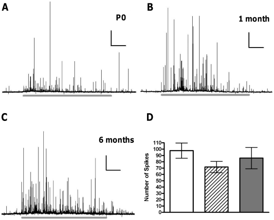
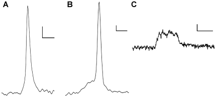
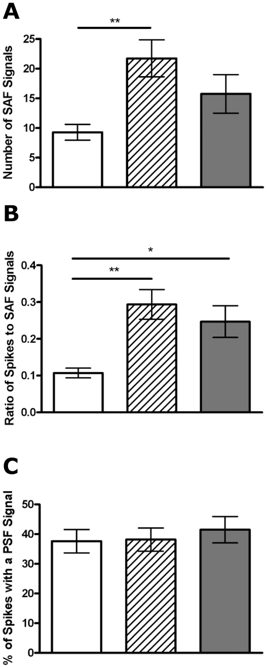
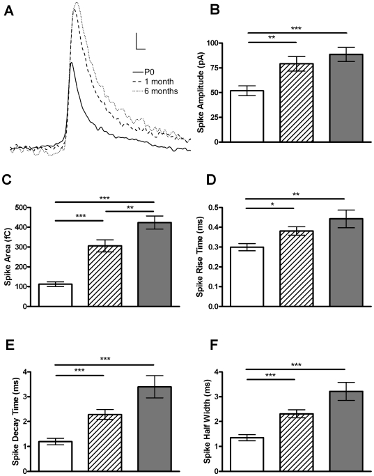
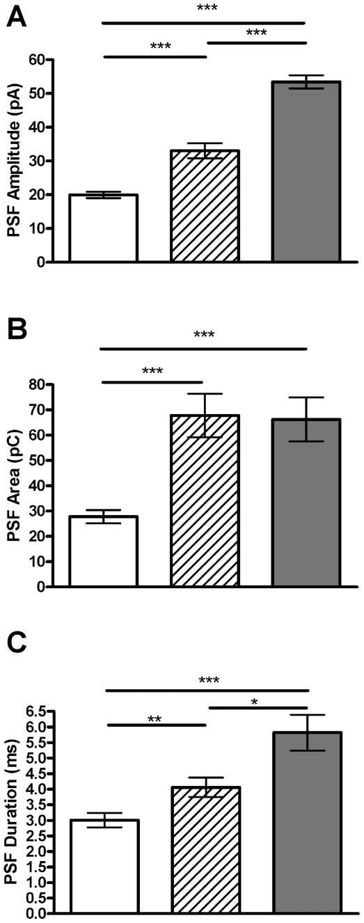
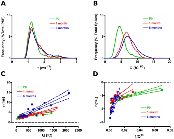
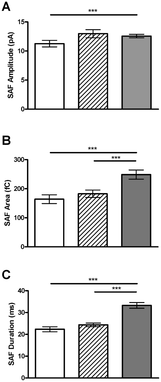
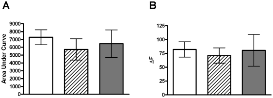
How fusion pore formation during exocytosis affects the subsequent release of vesicle contents remains incompletely understood. It is unclear if the amount released per vesicle is dependent upon the nature of the developing fusion pore and whether full fusion and transient kiss and run exocytosis are regulated by similar mechanisms. We hypothesise that if consistent relationships exist between these aspects of exocytosis then they will remain constant across any age. Using amperometry in mouse chromaffin cells we measured catecholamine efflux during single exocytotic events at P0, 1 month and 6 months. At all ages we observed full fusion (amperometric spike only), full fusion preceded by fusion pore flickering (pre-spike foot (PSF) signal followed by a spike) and pure "kiss and run" exocytosis (represented by stand alone foot (SAF) signals). We observe age-associated increases in the size of all 3 modes of fusion but these increases occur at different ages. The release probability of PSF signals or full spikes alone doesn't alter across any age in comparison with an age-dependent increase in the incidence of "kiss and run" type events. However, the most striking changes we observe are age-associated changes in the relationship between vesicle size and the membrane bending energy required for exocytosis. Our data illustrates that vesicle size does not regulate release probability, as has been suggested, that membrane elasticity or flexural rigidity change with age and that the mechanisms controlling full fusion may differ from those controlling "kiss and run" fusion.


Conflict of interest statement

Competing Interests: DJK has a nonfinancial interest as an Academic Editor of PLoS ONE.

Figures

Figure 1
Figure 1. Aging does not affect the number of vesicles undergoing full fusion.
Representative amperometric traces show the 60 second stimulation period (grey line below trace) in cells from (A) P0, (B) 1 month and (C) 6 months. The average number of events for each cell (D) illustrates the number of exocytotic events does not change with age. n = 14, 15 and 16 for P0 (white bar), 1 month (striped bar) and 6 months (grey bar), respectively. Scale bars in (A–C) represent 10sec and 100 pA.
Figure 2
Figure 2. Different types of fusion as observed using carbon fibre amperometry.
A full fusion event without a pre-spike foot signal (A), a full fusion event with a pre-spike foot signal (B) and a kiss and run event represented as a stand-alone foot signal (C) are shown. Scale bars represent 2 ms and 10 pA.
Figure 3
Figure 3. Aging affects the incidence of pre-spike foot signals and stand-alone foot signal events differently.
The number of stand-alone foot (SAF) signals observed increases with age (A) as does the ratio of SAF signals to full spikes (B), while the percentage of spike displaying a pre-spike foot (PSF) signal is maintained across all ages studied (C). n = 14, 15 and 16 cells for P0 (white bar), 1 month (striped bar) and 6 month (grey bar), respectively. *, p<0.05, **, p<0.01, calculated by Mann-Whitney U tests.
Figure 4
Figure 4. Aging alters the kinetics of full fusion.
(A) Examples of a typical amperometric spike at P0, 1 month and 6 months. Increases in spike amplitude (B), area (C), rise time (D), decay time (E) and half-width (F) are all evident, indicating that release kinetics and the amount released from each vesicle increases with age. Graphs represent the mean of each cells median value ± SEM. *, p<0.05; **, p<0.01; ***, p<0.001, calculated by Mann-Whitney U tests. n = 14, 15 and 16 for P0 (white bar), 1 month (striped bar) and 6 month (grey bar), respectively. Scale bars in (A) represent 10 ms and 10 pA.
Figure 5
Figure 5. The kinetics of the transient fusion pore increase with age.
The amplitude (A), area (B) and duration (C) of the pre-spike foot (PSF) signal change with aging. Graphs show means of all PSF signals at each age tested ± SEM. *, p<0.05; **, p<0.01; ***, p<0.001, calculated by Mann-Whitney U tests. n = 14, 15 and 16 cells and 466, 268, 599 events for P0 (white bar), 1 month (striped bar) and 6 month (grey bar), respectively.
Figure 6
Figure 6. Aging differentially affects membrane bending properties during exocytosis.
Frequency distribution of the cube root of pre-spike foot (PSF) signal duration (A) and spike area (B) illustrates the different ages at which these change. Correlations between PSF signal duration, τ, and spike area, Q (C) shows a linear relationship at all ages (P0: R2 = 0.64, p<0.01, 1 month: R2 = 0.82, p<0.0001, 6 months: R2 = 0.79, p<0.0001). Transforms of this data to ln(1/τ) vs 1/Q1/3 (D) also provides linear relationships at all ages (P0: R2 = 0.81, p<0.0001, 1 month: R2 = 0.44, p<0.05, 6 months: R2 = 0.59, p<0.01). The slope of this plot, representing membrane curvature , increases significantly with age. Green – P0, red – 1 month, blue – 6 months.
Figure 7
Figure 7. The kinetics of kiss and run fusion events changes with aging.
The amplitude (A), area (B) and duration (C) of stand-alone foot (SAF) signals all change with aging. Graphs show means of all SAF signals at each age tested ± SEM. ***, p<0.001, calculated by Mann-Whitney U tests. n = 14, 15 and 16 cells and 130, 273 and 252 events for P0 (white bar), 1 month (striped bar) and 6 month (grey bar), respectively.
Figure 8
Figure 8. Ca2+ entry is not altered with age in mouse chromaffin cells.
Cells from each age were loaded with the Ca2+-sensitive dye, Fluo-4, and stimulated for 60 sec with 70 mM K+ solution. The increase in fluorescence reflects Ca2+ entry and we find that the total increase in fluorescence (area under the curve; A) and the peak fluorescence change (ΔF; B) are unchanged at any age. Data is mean ± SEM, significance tested using one-way ANOVA. n = 16, 9 and 8 cells from P0 (white bar), 1 month (striped bar) and 6 month (grey bar), respectively. Cells were obtained from >3 mice in each age group.

Similar articles

Cited by

References

    1. Zhang Z, Jackson MB. Membrane bending energy and fusion pore kinetics in Ca(2+)-triggered exocytosis. Biophys J. 2010;98:2524–2534. - PMC - PubMed
    1. Zhou Z, Misler S, Chow RH. Rapid fluctuations in transmitter release from single vesicles in bovine adrenal chromaffin cells. Biophys J. 1996;70:1543–1552. - PMC - PubMed
    1. Amatore C, Arbault S, Bonifas I, Guille M. Quantitative investigations of amperometric spike feet suggest different controlling factors of the fusion pore in exocytosis at chromaffin cells. Biophys Chem. 2009;143:124–131. - PubMed
    1. Pothos EN, Mosharov E, Liu KP, Setlik W, Haburcak M, et al. Stimulation-dependent regulation of the pH, volume and quantal size of bovine and rodent secretory vesicles. J Physiol. 2002;542:453–476. - PMC - PubMed
    1. Amatore C, Arbault S, Bonifas I, Bouret Y, Erard M, et al. Correlation between vesicle quantal size and fusion pore release in chromaffin cell exocytosis. Biophys J. 2005;88:4411–4420. - PMC - PubMed

Publication types