. 2011 Nov 29;8(1):31.
doi: 10.1186/1742-9994-8-31.

An Intermediate in the evolution of superfast sonic muscles

Affiliations

An Intermediate in the evolution of superfast sonic muscles

Hin-Kiu Mok et al. Front Zool. .

Abstract

Background: Intermediate forms in the evolution of new adaptations such as transitions from water to land and the evolution of flight are often poorly understood. Similarly, the evolution of superfast sonic muscles in fishes, often considered the fastest muscles in vertebrates, has been a mystery because slow bladder movement does not generate sound. Slow muscles that stretch the swimbladder and then produce sound during recoil have recently been discovered in ophidiiform fishes. Here we describe the disturbance call (produced when fish are held) and sonic mechanism in an unrelated perciform pearl perch (Glaucosomatidae) that represents an intermediate condition in the evolution of super-fast sonic muscles.

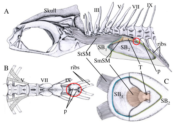
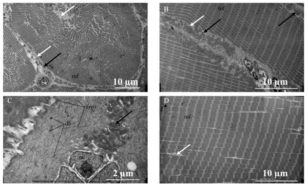
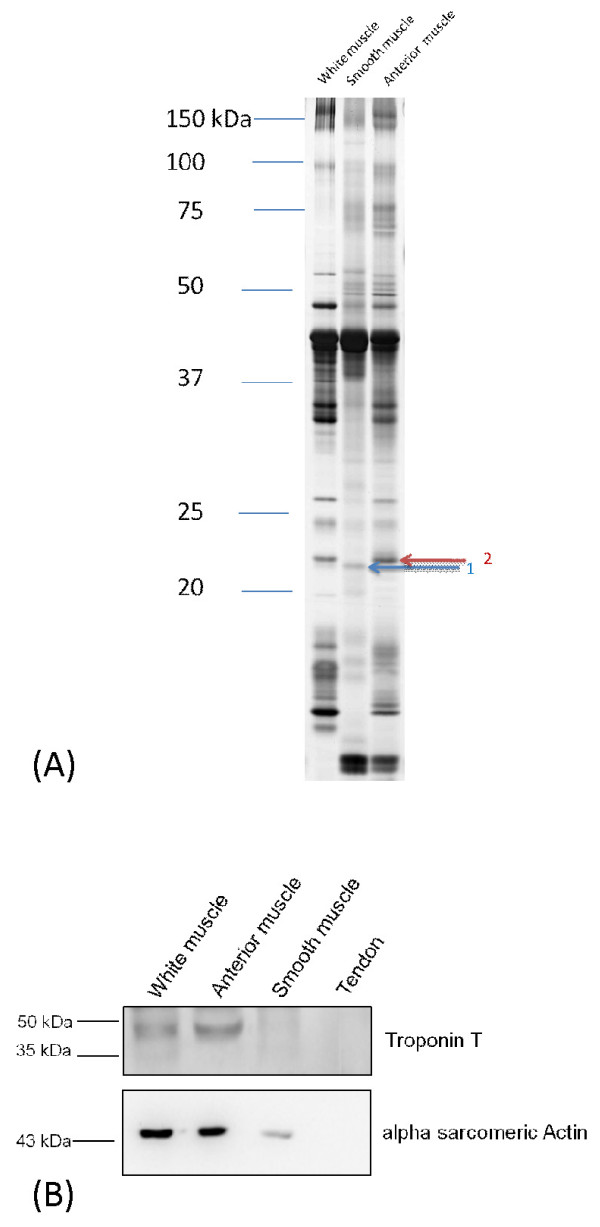
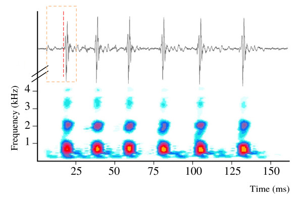
Results: The pearl perch disturbance call is a two-part sound produced by a fast sonic muscle that rapidly stretches the bladder and an antagonistic tendon-smooth muscle combination (part 1) causing the tendon and bladder to snap back (part 2) generating a higher-frequency and greater-amplitude pulse. The smooth muscle is confirmed by electron microscopy and protein analysis. To our knowledge smooth muscle attachment to a tendon is unknown in animals.

Conclusion: The pearl perch, an advanced perciform teleost unrelated to ophidiiform fishes, uses a slow type mechanism to produce the major portion of the sound pulse during recoil, but the swimbladder is stretched by a fast muscle. Similarities between the two unrelated lineages, suggest independent and convergent evolution of sonic muscles and indicate intermediate forms in the evolution of superfast muscles.


Figures

Figure 1
Figure 1
Drawings of the sonic system of Glaucosoma buergeri. A. Lateral view of the sonic system. Latin numbers designate individual vertebra. B. Ventral view of the vertebral column illustrating processes that support the posterior swimbladder and attachment of the sonic tendon. The red circle indicates the insertion of the tendon on the paraphoyses of vertebra IX. C. Ventral view of the dorsal wall of the swimbladder (base of the swimbladder removed) illustrating the attachment of the tendon-smooth muscle to the inner tunica externa. Abbreviations: p parapohysis of vertebra IX, SB1 anterior part of the swimbladder, SB2 posterior part of the swimbladder, StSM: anterior striated sonic muscle, SmSM: smooth sonic muscle, and T tendon from vertebra IX to the smooth sonic muscle. The swimbladder fenestra, although not visible because it is covered by the tunica externa, is situated between SB1 and SB2.
Figure 2
Figure 2
Electron micrographs of sonic and hypaxial muscles of the pearl perch Glaucosoma buergeri. A. Cross section of a sonic fiber from the anterior-striated sonic muscle. B. Longitudinal section of a fiber from the anterior-striated sonic muscle. C. Longitudinal section of a fiber from the smooth muscle. D. Longitudinal section of epaxial trunk muscle. Note thicker myofibrils and thinner sarcoplasmic reticulum compared to sonic fibers. Abbreviations: bv blood vessel, db dense bodies, mf myofibrils, myo myofilaments in the smooth muscle, nu nucleus. Dark arrows indicate mitochondria and white arrows sarcoplasmic reticulum.
Figure 3
Figure 3
SDS-PAGE (A) and Western blotting (B) analyses of Glaucosoma buergeri muscles. Band 1, blue arrow: a protein homologous to the smooth-muscle specific protein transgelin, identified using LC-MS/MS followed by a database search, was revealed only in the smooth muscle. Band 2 red arrow: indicates myosin light chain 3 (see Additional File 2) from striated muscle (trunk white and anterior sonic muscle). B. Troponin T (upper panel) present in striated but not in the internal smooth muscle and alpha sacromeric actin (lower panel) present in muscles but not in the tendon. Actin, as expected, is expressed at a lower level in smooth than in striated muscles.
Figure 4
Figure 4
Oscillogram and sonagram of a series of sound pulses evoked by touching the abdomen of the pearl perch Glaucosoma buergeri. The box in the oscillogram designates the first pulse, and the vertical dashed line separates pulse parts 1 and 2.

References

    1. Bass AH, Gilland EH, Baker R. Evolutionary Origins for Social Vocalization in a Vertebrate Hindbrain-Spinal Compartment. Science. 2008;321:417–421. doi: 10.1126/science.1157632. - DOI - PMC - PubMed
    1. Ladich F, Fine M. In: Communication in Fishes. Ladich F, Collin SP, Moller P, Kapoor BG, editor. Vol. 1. Endfield: Science Publishers; 2006. Sound-Generating Mechanisms in Fishes: A Unique Diversity in Vertebrates; pp. 3–43.
    1. Parmentier E, Diogo R. In: Communication in Fishes. Ladich F, Collin SP, Moller P, Kapoor BG, editor. Enfield, NH, USA: Jersey Plymouth: Science Publishers; 2006. Evolutionary trends of swimbladder sound mechanisms in some teleost fishes; pp. 43–68.
    1. Bass AH, McKibben JR. Neural mechanisms and behaviors for acoustic communication in teleost fish. Progress in Neurobiology. 2003;69:1–26. doi: 10.1016/S0301-0082(03)00004-2. - DOI - PubMed
    1. Fine ML, Thorson RF. Use of Passive Acoustics for Assessing Behavioral Interactions in Individual Toadfish. Transactions of the American Fisheries Society. 2008;137:627–637. doi: 10.1577/T04-134.1. - DOI