

Evaluation of multiple-atlas-based strategies for segmentation of the thyroid gland in head and neck CT images for IMRT
- PMID: 22126838
- PMCID: PMC3505993
- DOI: 10.1088/0031-9155/57/1/93
Evaluation of multiple-atlas-based strategies for segmentation of the thyroid gland in head and neck CT images for IMRT
Erratum in
- Phys Med Biol. 2012 Apr 7;57(7):2101
Abstract
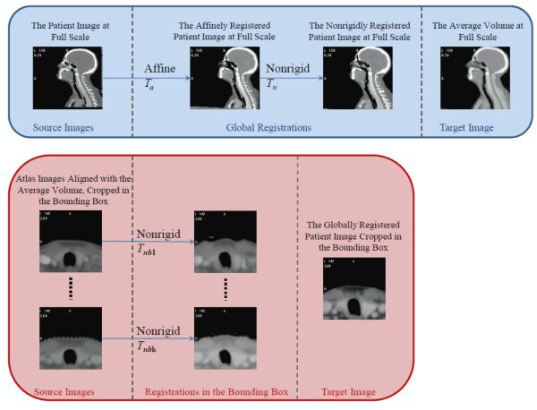
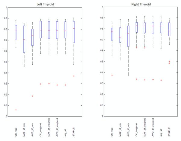
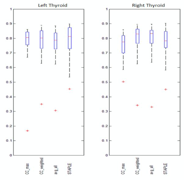
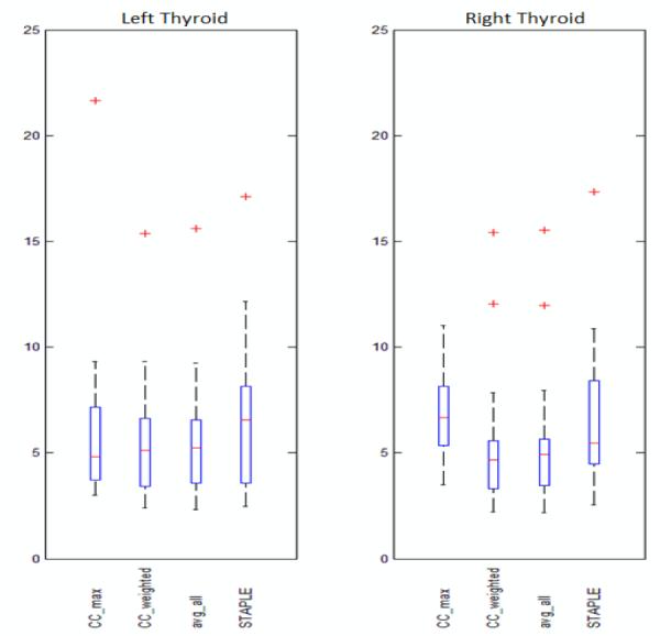
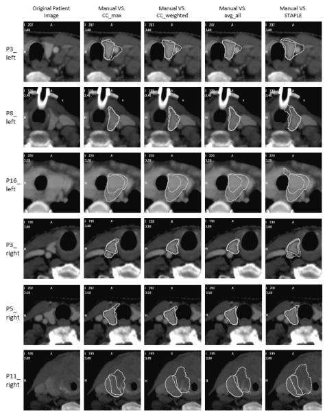
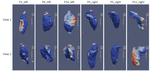
Segmenting the thyroid gland in head and neck CT images is of vital clinical significance in designing intensity-modulated radiation therapy (IMRT) treatment plans. In this work, we evaluate and compare several multiple-atlas-based methods to segment this structure. Using the most robust method, we generate automatic segmentations for the thyroid gland and study their clinical applicability. The various methods we evaluate range from selecting a single atlas based on one of three similarity measures, to combining the segmentation results obtained with several atlases and weighting their contribution using techniques including a simple majority vote rule, a technique called STAPLE that is widely used in the medical imaging literature, and the similarity between the atlas and the volume to be segmented. We show that the best results are obtained when several atlases are combined and their contributions are weighted with a measure of similarity between each atlas and the volume to be segmented. We also show that with our data set, STAPLE does not always lead to the best results. Automatic segmentations generated by the combination method using the correlation coefficient (CC) between the deformed atlas and the patient volume, which is the most accurate and robust method we evaluated, are presented to a physician as 2D contours and modified to meet clinical requirements. It is shown that about 40% of the contours of the left thyroid and about 42% of the right thyroid can be used directly. An additional 21% on the left and 24% on the right require only minimal modification. The amount and the location of the modifications are qualitatively and quantitatively assessed. We demonstrate that, although challenged by large inter-subject anatomical discrepancy, atlas-based segmentation of the thyroid gland in IMRT CT images is feasible by involving multiple atlases. The results show that a weighted combination of segmentations by atlases using the CC as the similarity measure slightly outperforms standard combination methods, e.g. the majority vote rule and STAPLE, as well as methods selecting a single most similar atlas. The results we have obtained suggest that using our contours as initial contours to be edited has clinical value.
Figures
Similar articles
-
Atlas ranking and selection for automatic segmentation of the esophagus from CT scans.Phys Med Biol. 2017 Nov 14;62(23):9140-9158. doi: 10.1088/1361-6560/aa94ba. Phys Med Biol. 2017. PMID: 29049027 Free PMC article.
-
Automatic segmentation of head and neck CT images for radiotherapy treatment planning using multiple atlases, statistical appearance models, and geodesic active contours.Med Phys. 2014 May;41(5):051910. doi: 10.1118/1.4871623. Med Phys. 2014. PMID: 24784389 Free PMC article.
-
Clinical evaluation of multi-atlas based segmentation of lymph node regions in head and neck and prostate cancer patients.Radiat Oncol. 2013 Oct 3;8:229. doi: 10.1186/1748-717X-8-229. Radiat Oncol. 2013. PMID: 24090107 Free PMC article.
-
Auto-segmentation of low-risk clinical target volume for head and neck radiation therapy.Pract Radiat Oncol. 2014 Jan-Feb;4(1):e31-7. doi: 10.1016/j.prro.2013.03.003. Epub 2013 May 3. Pract Radiat Oncol. 2014. PMID: 24621429
-
Thyroid Screening Techniques via Bioelectromagnetic Sensing: Imaging Models and Analytical and Computational Methods.Sensors (Basel). 2024 Sep 21;24(18):6104. doi: 10.3390/s24186104. Sensors (Basel). 2024. PMID: 39338849 Free PMC article. Review.
Cited by
-
Atlas ranking and selection for automatic segmentation of the esophagus from CT scans.Phys Med Biol. 2017 Nov 14;62(23):9140-9158. doi: 10.1088/1361-6560/aa94ba. Phys Med Biol. 2017. PMID: 29049027 Free PMC article.
-
Hierarchical performance estimation in the statistical label fusion framework.Med Image Anal. 2014 Oct;18(7):1070-81. doi: 10.1016/j.media.2014.06.005. Epub 2014 Jul 4. Med Image Anal. 2014. PMID: 25033470 Free PMC article.
-
Validation and Modification of a Prediction Model for Acute Cardiac Events in Patients With Breast Cancer Treated With Radiotherapy Based on Three-Dimensional Dose Distributions to Cardiac Substructures.J Clin Oncol. 2017 Apr 10;35(11):1171-1178. doi: 10.1200/JCO.2016.69.8480. Epub 2017 Jan 17. J Clin Oncol. 2017. PMID: 28095159 Free PMC article.
-
Automated segmentation of the thyroid gland on thoracic CT scans by multiatlas label fusion and random forest classification.J Med Imaging (Bellingham). 2015 Oct;2(4):044006. doi: 10.1117/1.JMI.2.4.044006. Epub 2015 Dec 30. J Med Imaging (Bellingham). 2015. PMID: 26730397 Free PMC article.
-
Evaluation of Multi-Atlas Label Fusion for In Vivo MRI Orbital Segmentation.J Med Imaging (Bellingham). 2014 Jul 18;1(2):024002. doi: 10.1117/1.JMI.1.2.024002. J Med Imaging (Bellingham). 2014. PMID: 25558466 Free PMC article.
References
-
- Aljabar P, Heckermann RA, Hammers A, Hajnal JV, Rueckert D. Multi-atlas based segmentation of brain images: Atlas selection and its effect on accuracy. NeuroImage. 2009;46:726–38. - PubMed
-
- Alterio D, Jereczek-Fossa BA, Franchi B, et al. Thyroid disorder in patients treated with radiotherapy for head-and-neck cancer: A retrospective analysis of seventy-three patients. Int J Radiat Oncol Biol Phys. 2007;67:144–50. - PubMed
-
- Artaechevarria X, Munoz-Barrutia A, Ortiz-de-Solorzano C. Combination strategies in multi-atlas image segmentation: application to brain MR data. IEEE Trans. on Med. Imag. 2009;28:1266–77. - PubMed
-
- Bethge W, Guggenberger D, Bamberg M, et al. Thyroid toxicity of treatment for Hodgkin’s disease. Ann Hematol. 2000;79:114–8. - PubMed
-
- Bhandare N, Kennedy L, Malyapa RS, et al. Primary and central hypothyroidism after radiotherapy for head-and-neck tumors. Int J Radiat Oncol Biol Phys. 2007;68:1131–9. - PubMed