Review
. 2012 Jan;177(1):24-31.
doi: 10.1016/j.jsb.2011.11.007. Epub 2011 Nov 22.

Nuclear lamina at the crossroads of the cytoplasm and nucleus

Affiliations
Review

Nuclear lamina at the crossroads of the cytoplasm and nucleus

Larry Gerace et al. J Struct Biol. 2012 Jan.

Abstract

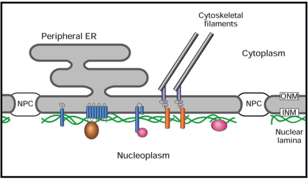
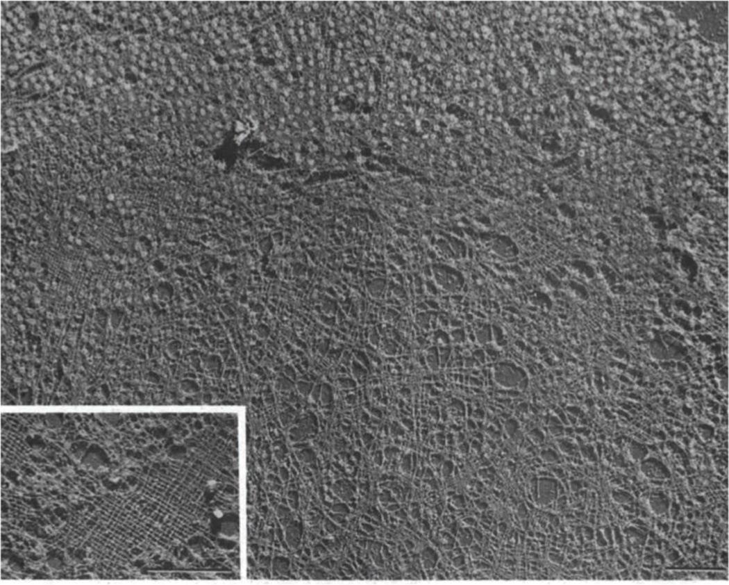
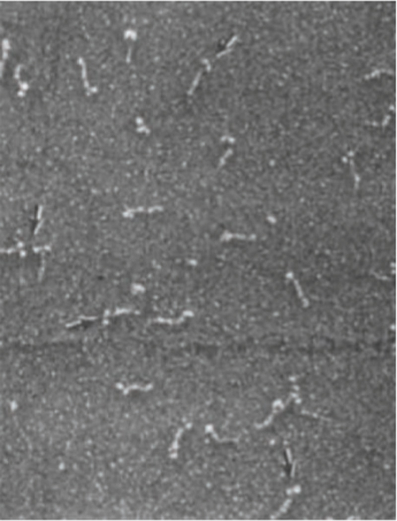
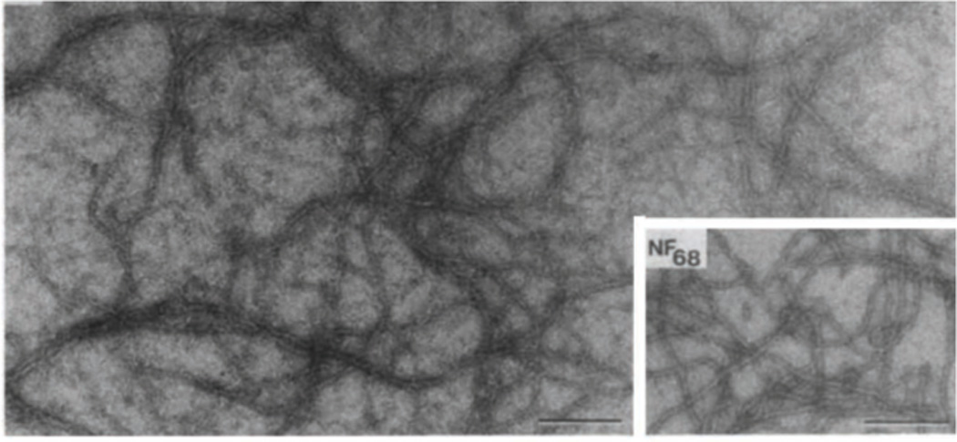
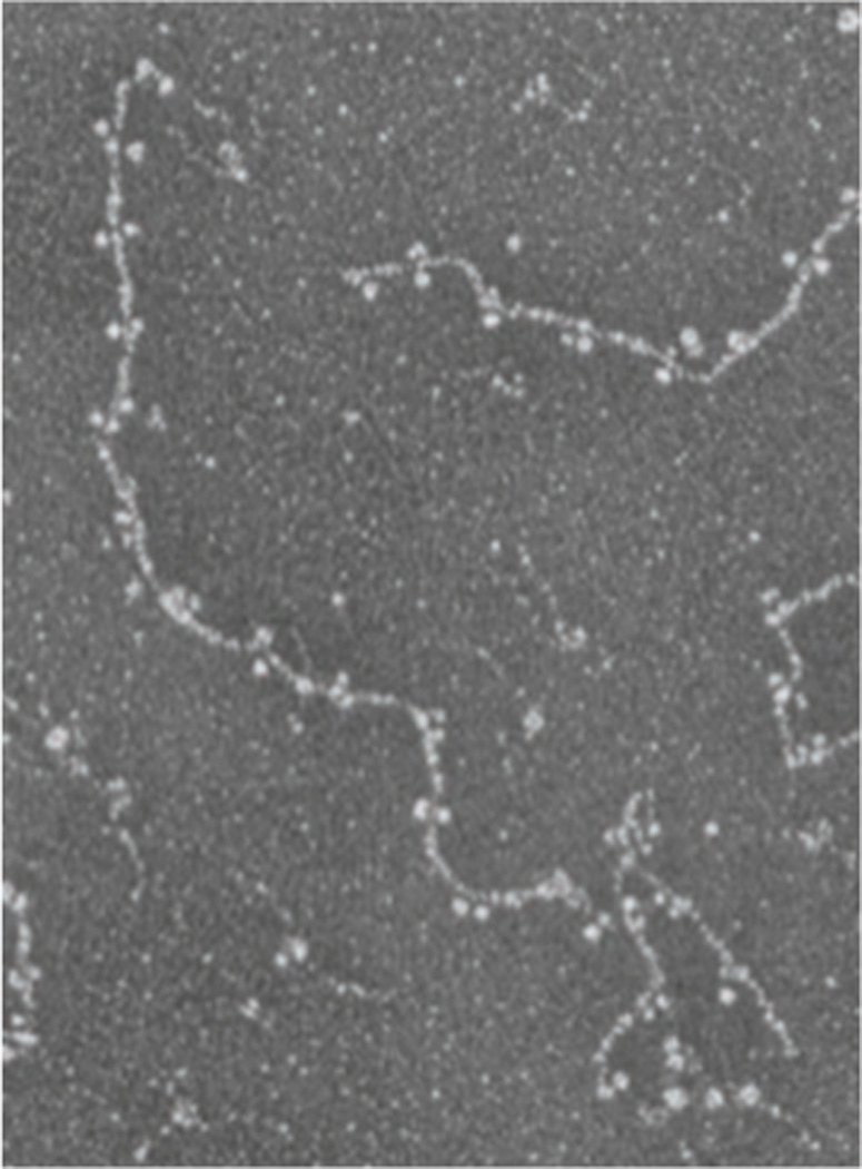
The nuclear lamina is a protein meshwork that lines the nuclear envelope in metazoan cells. It is composed largely of a polymeric assembly of lamins, which comprise a distinct sequence homology class of the intermediate filament protein family. On the basis of its structural properties, the lamina originally was proposed to provide scaffolding for the nuclear envelope and to promote anchoring of chromatin and nuclear pore complexes at the nuclear surface. This viewpoint has expanded greatly during the past 25 years, with a host of surprising new insights on lamina structure, molecular composition and functional attributes. It has been established that the self-assembly properties of lamins are very similar to those of cytoplasmic intermediate filament proteins, and that the lamin polymer is physically associated with components of the cytoplasmic cytoskeleton and with a multitude of chromatin and inner nuclear membrane proteins. Cumulative evidence points to an important role for the lamina in regulating signaling and gene activity, and in mechanically coupling the cytoplasmic cytoskeleton to the nucleus. The significance of the lamina has been vaulted to the forefront by the discovery that mutations in lamins and lamina-associated polypeptides lead to an array of human diseases. A key future challenge is to understand how the lamina integrates pathways for mechanics and signaling at the molecular level. Understanding the structure of the lamina from the atomic to supramolecular levels will be essential for achieving this goal.


Figures

Figure 1
Figure 1. Schematic diagram of the nuclear envelope
The outer nuclear membrane (ONM), which is continuous with the peripheral ER, is joined to inner nuclear membrane (INM) at the nuclear pore complex (NPC). The nuclear lamina comprises lamin filament polymers (green) and associated membrane-spanning proteins (blue), and peripheral proteins (brown and pink). The lamina is connected to the cytoplasmic cytoskeletal filaments by the LINC complex, which consists of Sun domain proteins (orange) spanning the INM that attached to nesprins (grey) that span the ONM.
Figure 2
Figure 2. View of the nuclear lamina from Xenopus oocyte en face
Electron micrograph of a replica of a freeze dried/ metal shadowed Xenopus oocyte NE after treatment with Triton X-100. The lamina is revealed as a quasi-orthogonal filament meshwork in areas where NPCs have been removed by mechanical forces (inset), but the network also can be seen in NPC-attached regions (upper left). Reproduced from (Aebi et al., 1986) with permission. Bars, 1 µm.
Figure 3
Figure 3. Structure of the purified lamins A/C dimers by rotary shadowing
Electron micrograph of a sample of purified lamins A/C from rat liver NEs that was prepared by glycerol spraying/ rotary shadowing. The lamin dimer is seen as an ~52 nm rod attached to 2 masses (arrows). Reproduced from (Aebi et al., 1986) with permission.
Figure 4
Figure 4. Reconstitution of 10 nm filaments from purified lamins A/C
Electron micrograph of a negatively stained specimen of rat liver lamins A/C dialyzed into 25 mM MES, 200 mM NaC1 pH 6.5. Inset. For comparison, a sample of 10 nm filaments reconstituted from the purified 68 kD neurofilament protein of bovine brain is shown. Bars, 250 nm. Reproduced from (Aebi et al., 1986) with permission.
Figure 5
Figure 5. In vitro assembly of head-to-tail dimers chicken lamin B2
Electron micrograph of a glycerol sprayed/ rotary shadowed sample of purified recombinant chicken lamin B2 dimers dialyzed into a buffer containing 25 mM MES, 150 mM NaC1 pH 6.5. Longitudinal head-to-tail polymers of lamin dimers are shown. ©1991 Rockefeller University Press. Originally published in J. Cell Biol. 113:485–495.

References

    1. Aaronson RP, Blobel G. On the attachment of the nuclear pore complex. J Cell Biol. 1974;62:746–754. - PMC - PubMed
    1. Aaronson RP, Blobel G. Isolation of nuclear pore complexes in association with a lamina. Proc Natl Acad Sci U S A. 1975;72:1007–1011. - PMC - PubMed
    1. Aebi U, Cohn J, Buhle L, Gerace L. The nuclear lamina is a meshwork of intermediate-type filaments. Nature. 1986;323:560–564. - PubMed
    1. Anderson DJ, Vargas JD, Hsiao JP, Hetzer MW. Recruitment of functionally distinct membrane proteins to chromatin mediates nuclear envelope formation in vivo. J Cell Biol. 2009;186:183–191. - PMC - PubMed
    1. Ben-Harush K, Wiesel N, Frenkiel-Krispin D, Moeller D, Soreq E, et al. The supramolecular organization of the C. elegans nuclear lamin filament. J Mol Biol. 2009;386:1392–1402. - PubMed

Publication types