Review
. 2011 Nov 30;13(1):22-37.
doi: 10.1038/nrn3138.

The stressed synapse: the impact of stress and glucocorticoids on glutamate transmission

Affiliations
Review

The stressed synapse: the impact of stress and glucocorticoids on glutamate transmission

Maurizio Popoli et al. Nat Rev Neurosci. .

Abstract

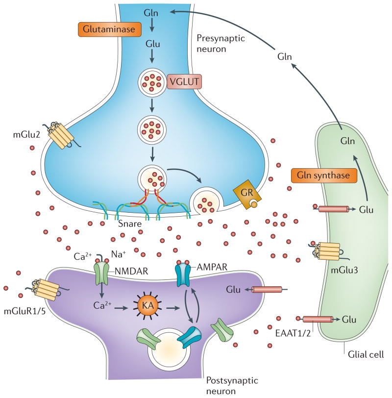
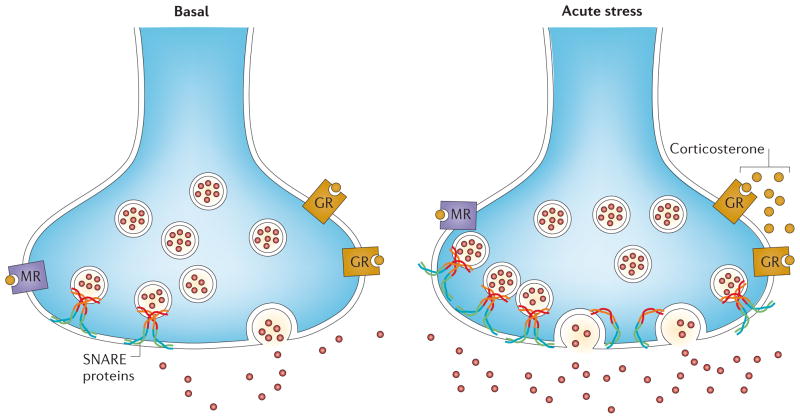
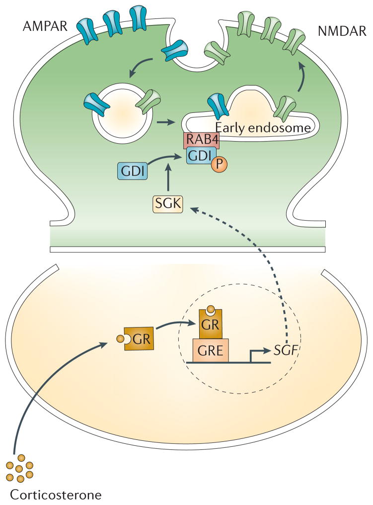
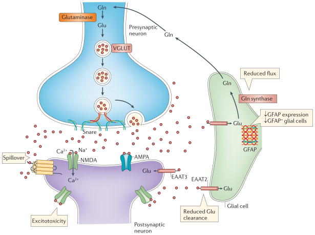
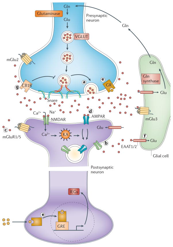
Mounting evidence suggests that acute and chronic stress, especially the stress-induced release of glucocorticoids, induces changes in glutamate neurotransmission in the prefrontal cortex and the hippocampus, thereby influencing some aspects of cognitive processing. In addition, dysfunction of glutamatergic neurotransmission is increasingly considered to be a core feature of stress-related mental illnesses. Recent studies have shed light on the mechanisms by which stress and glucocorticoids affect glutamate transmission, including effects on glutamate release, glutamate receptors and glutamate clearance and metabolism. This new understanding provides insights into normal brain functioning, as well as the pathophysiology and potential new treatments of stress-related neuropsychiatric disorders.


Conflict of interest statement

Competing financial interests

MP: Dr. Popoli received support and/or has consulted for Abiogen, GlaxoSmith-Kline, MerckSharp and Dohme, Servier and Fidia

ZY: Reports no competing financial interests

BMc: Reports no competing financial interests

GS: Dr. Sanacora has received consulting fees form AstraZeneca, Avanier Pharmaceuticals, Bristol-Myers Squibb, Evotec, Eli Lilly & Co., Hoffman La-Roche, Johnson & Johnson, Novartis, and Novum Pharmaceuticals over the last 24 months. He has also received additional grant support from AstraZeneca, Bristol-Myers Squibb, Hoffman La-Roche, Merck & Co., and Sepracor Inc over the last 24 months. In addition he is a co-inventor on filed patent application by Yale University (PCTWO06108055A1).

Figures

Figure 1
Figure 1. The tripartite glutamate synapse
Neuronal glutamate is both synthesized de novo from glucose (not shown) and from glutamine (Gln) supplied by glial cells. Glutamate is then packaged into synaptic vesicles by vesicular glutamate transporters (vGluT). SNARE complex proteins mediate the interaction and fusion of vesicles with the presynaptic membrane. After release into the extracellular space, glutamate binds to ionotropic (NMDA, AMPA) and metabotropic (mGluR 1–8) receptors on the membranes of both post-synaptic and pre-synaptic neurons and glial cells. Upon binding, the receptors initiate various responses, including membrane depolarization, activation of intracellular messenger cascades, modulation of local protein synthesis and, eventually, gene expression (not shown). Surface expression and function of NMDARs and AMPARs is dynamically regulated by protein synthesis and degradation and receptor trafficking between the postsynaptic membrane and endosomes. The insertion and removal of postsynaptic receptors provide a mechanism for long-term modulation of synaptic strength. Glutamate is cleared from the synapse through excitatory amino acid transporters (EAATs) on neighbouring glial cells (EAAT1 and 2) and, to a lesser extent, on neurons (EAAT 3–5). Within the glial cell, glutamate is converted to glutamine by glutamine synthetase and the glutamine is subsequently released via system N transporters and taken up by neurons through System A sodium-coupled amino acid transporters to complete the glutamate–glutamine cycle.
Figure 2
Figure 2. Acute stress rapidly enhances glutamate release in prefrontal/frontal cortex
Acute footshock stress enhances depolarization-evoked release of glutamate from presynaptic terminals of rat prefrontal/frontal cortex. The acute stress response involves a rapid increase of circulating levels of corticosterone, which binds to membrane-located glucocorticoid receptors (GRs). This induces a rapid GR-mediated increase of presynaptic SNARE protein complexes (which mediate fusion of synaptic vesicles) in the presynaptic membrane. Because the number of SNARE complexes per vesicle is reputed to be constant, this suggests that acute stress induces an increase of the readily releasable pool of glutamate vesicles. The signalling pathways downstream of glucocorticoid receptor activation that induce the increase of the readily releasable pool are unknown.
Figure 3
Figure 3. Stress induces changes in glutamate receptor trafficking and function in the prefrontal cortex
In response to acute stress, activation of glucocorticoid receptors (GRs) triggers the upregulation of transcription of the gene encoding serum- and glucocorticoid-inducible kinase (SGK) 1/3. SGK1/3 phosphorylates GDP dissociation inhibitor (GDI) and thereby increases the formation of GDI-Rab4 complexes. Consequently, Rab4-mediated recycling of NMDARs and AMPARs from early endosomes (EE) to the plasma membrane is enhanced, and this results in increased glutamate receptor expression at the synaptic membrane and potentiated glutamatergic transmission,.
Figure 4
Figure 4. Chronic stress affects glial cells and glutamate metabolism
Accumulating evidence suggests that chronic stress has significant effects on glial cell function. Several studies have demonstrated decreases in the expression of glial fibrillary acid protein (GFAP) and in the number of GFAP-expressing glial cells in the hippocampus and PFC following exposure to chronic stress. Chronic stress may also impair the ability to effectively clear synaptic glutamate through glial excitatory amino acid transporters (EAATs). This may lead to glutamate spillover and, ultimately, increased activation of extrasynaptic glutamate receptors resulting in excitotoxicity, a process that has been proposed to occur in several neurodegenerative disorders, and possibly after exposure to chronic stress. Finally, chronic stress may decrease the rates of flux through the glutamate–glutamine cycle, resulting in reduced glutamate metabolism.
Figure 5
Figure 5. Pharmacological targets
Observations of stress-induced effects on the glutamate synapse have suggested several unique forms of pharmacological interventions for stress related disorders such as mood and anxiety disorders. Drugs that modify glutamate release (a), such as lamotrigine and rilzuole, have been shown to have antidepressant-like actions in rodent models and in clinical trials,,. In addition, negative and positive allosteric modulators of group II mGluRs that also modulate presynaptic glutamate release (not shown), have been shown to have antidepressant-like actions in rodent models. Drugs targeting NMDA receptors (b), especially NMDA antagonists (ketamine, RO 25-6981, and CP101,606) have demonstrated rapid and robust antidepressant-like effects in both rodent models and clinical trials,. Positive and negative allosteric modulators of the mGlu 5 receptor (c) have been shown to possess antidepressant and anxiolytic properties in preclinical studies Drugs targeting AMPA receptors (d), especially agents that potentiate the activation of AMPA receptors, have both nootropic (cognition-enhancing) properties and antidepressant-like effects in rodent models. Various agents that regulate glucocorticoid signalling have effects on memory and possess mood and anxiety modifying properties (e). Drugs such as riluzole and ceftriaxone that indirectly facilitate glutamate transport into glia (f), possess both neuroprotective and antidepressant-like effects,,. Considering endocannabinoids are reduced in the PFC and hippocampus in animal models of depression, and CB1 receptor stimulation in the PFC and hippocampus is anxiolytic and antidepressant, targeted pharmacological augmentation of endocannabinoid signalling (g) has recently been proposed as a promising therapeutic strategy for the treatment of mood and anxiety disorders.

References

    1. Selye H. A syndrome produced by diverse nocuous agents. Nature. 1936;138:32. - PubMed
    1. Lazarus RS, Folkman S. Stress, Appraisal and Coping. Springer Verlag; New York: 1984.
    1. McEwen BS. Protective and damaging effects of stress mediators. New England Journal of Medicine. 1998;338:171–179. - PubMed
    1. McEwen BS, Gianaros PJ. Stress- and allostasis-induced brain plasticity. Annu Rev Med. 2011;62:431–445. doi: 10.1146/annurev-med-052209-100430. - DOI - PMC - PubMed
    1. Liston C, et al. Stress-induced alterations in prefrontal cortical dendritic morphology predict selective impairments in perceptual attentional set-shifting. J Neurosci. 2006;26:7870–7874. doi: 10.1523/JNEUROSCI.1184-06.2006. 26/30/7870 [pii] - DOI - PMC - PubMed

Publication types