

Retinoic acid signaling regulates sonic hedgehog and bone morphogenetic protein signalings during genital tubercle development
- PMID: 22127979
- PMCID: PMC5837284
- DOI: 10.1002/bdrb.20344
Retinoic acid signaling regulates sonic hedgehog and bone morphogenetic protein signalings during genital tubercle development
Abstract
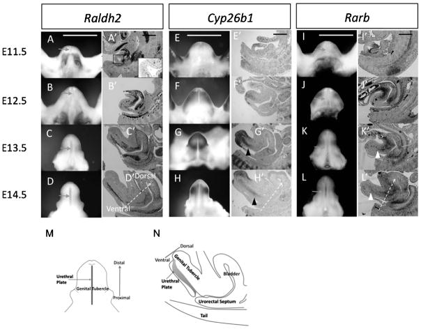
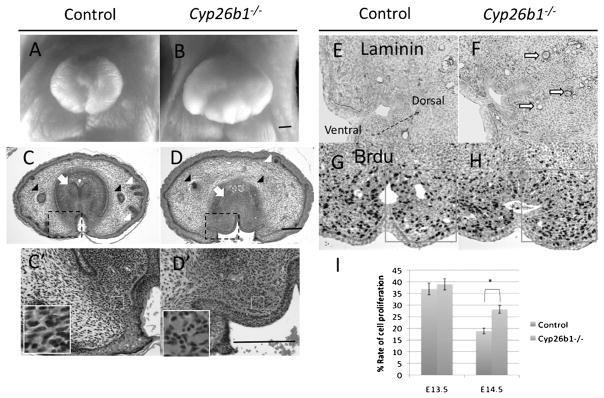
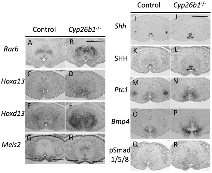
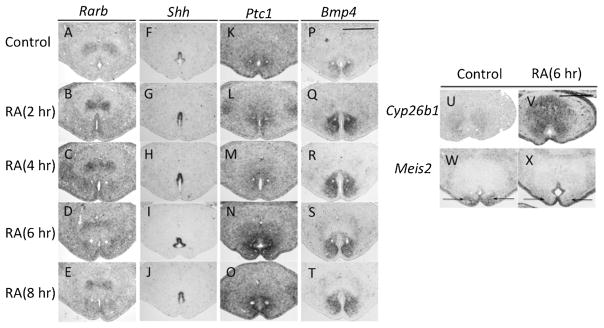
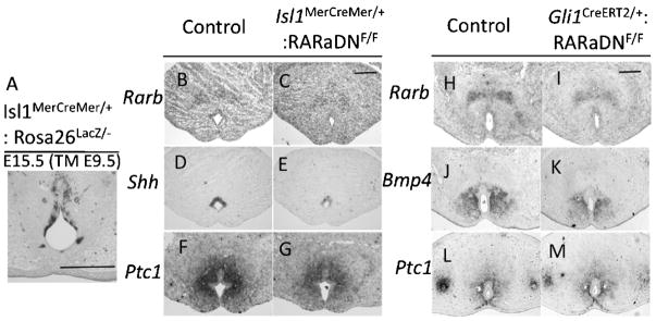
Retinoic acid (RA) plays pivotal roles in organogenesis, and both excessive and reduced amounts of RA cause developmental abnormalities. Reproductive organs are susceptible to teratogen toxigenicity, and the genital tubercle (GT) is one such representative organ. The physiological function of endogenous RA signaling and the mechanisms of RA-induced teratogenicity are poorly understood during the GT development. The objective of this study is to understand the developmental and teratogenic roles of RA during GT development by analyzing genetically modified mouse models. We found dynamic patterns of gene expression for the RA-synthesizing enzyme, Raldh2, and for the RA-catabolizing enzyme, Cyp26b1, during GT development. Rarb, an indicator gene for RA signaling, starts its expression in the prospective corpus cavernosum penis and in the urethral plate epithelium (UE), which plays central roles during GT development. Excessive RA signaling in Cyp26b1(-/-) mutants leads to abnormal extents of cell proliferation and differentiation during GT development, and also upregulates expression of growth factor signalings. They include Sonic hedgehog (Shh) signaling and Bone morphogenetic protein (Bmp) signaling, which are expressed in the UE and its bilateral mesenchyme. RA signaling positively regulatesShh and Bmp4 expression during GT development as testified also by the experiment of RA administration and analyses of loss-of-function of RA signaling mutants. Thus, RA signaling is involved in the developmental cascade necessary for UE formation and GT development.
Keywords: Bone morphogenetic protein (Bmp); Sonic hedgehog (Shh); genital tubercle; retinoic acid; urethral plate epithelium.
© 2011 Wiley-Liss, Inc.
Figures
Similar articles
-
Unique functions of Sonic hedgehog signaling during external genitalia development.Development. 2001 Nov;128(21):4241-50. doi: 10.1242/dev.128.21.4241. Development. 2001. PMID: 11684660
-
Bmp4 is an essential growth factor for the initiation of genital tubercle (GT) outgrowth.Congenit Anom (Kyoto). 2020 Jan;60(1):15-21. doi: 10.1111/cga.12326. Epub 2019 Feb 17. Congenit Anom (Kyoto). 2020. PMID: 30714224
-
Sonic hedgehog and bone morphogenetic protein 4 expressions in the hindgut region of murine embryos with anorectal malformations.J Pediatr Surg. 2004 Feb;39(2):170-3; discussion 170-3. doi: 10.1016/j.jpedsurg.2003.10.009. J Pediatr Surg. 2004. PMID: 14966734
-
Sonic hedgehog and bone morphogenetic protein-4 signaling pathway involved in epithelial cell renewal along the radial axis of the intestine.Digestion. 2008;77 Suppl 1:42-7. doi: 10.1159/000111487. Epub 2008 Jan 18. Digestion. 2008. PMID: 18204261 Review.
-
Development of the external genitalia: conserved and divergent mechanisms of appendage patterning.Dev Dyn. 2011 May;240(5):1108-15. doi: 10.1002/dvdy.22631. Epub 2011 Apr 4. Dev Dyn. 2011. PMID: 21465625 Free PMC article. Review.
Cited by
-
Exploration of the DARTable Genome- a Resource Enabling Data-Driven NAMs for Developmental and Reproductive Toxicity Prediction.Front Toxicol. 2022 Jan 19;3:806311. doi: 10.3389/ftox.2021.806311. eCollection 2021. Front Toxicol. 2022. PMID: 35295108 Free PMC article.
-
Retinoic acid signaling pathways in development and diseases.Bioorg Med Chem. 2014 Jan 15;22(2):673-83. doi: 10.1016/j.bmc.2013.11.025. Epub 2013 Nov 22. Bioorg Med Chem. 2014. PMID: 24393720 Free PMC article. Review.
-
Disruption of Retinol (Vitamin A) Signaling by Phthalate Esters: SAR and Mechanism Studies.PLoS One. 2016 Aug 17;11(8):e0161167. doi: 10.1371/journal.pone.0161167. eCollection 2016. PLoS One. 2016. PMID: 27532513 Free PMC article.
-
Replication of Caucasian loci associated with bone mineral density in Koreans.Osteoporos Int. 2013 Oct;24(10):2603-10. doi: 10.1007/s00198-013-2354-1. Epub 2013 Apr 11. Osteoporos Int. 2013. PMID: 23575750
-
Coordinated activity of Spry1 and Spry2 is required for normal development of the external genitalia.Dev Biol. 2014 Feb 1;386(1):1-11. doi: 10.1016/j.ydbio.2013.12.014. Epub 2013 Dec 18. Dev Biol. 2014. PMID: 24361260 Free PMC article.
References
-
- Ahn S, Joyner AL. Dynamic changes in the response of cells to positive hedgehog signaling during mouse limb patterning. Cell. 2004;118:505–516. - PubMed
-
- Batourina E, Choi C, Paragas N, Bello N, Hensle T, Costantini FD, Schuchardt A, Bacallao RL, Mendelsohn CL. Distal ureter morphogenesis depends on epithelial cell remodeling mediated by vitamin A and Ret. Nat Genet. 2002;32:109–115. - PubMed
-
- Chiu HS, Szucsik JC, Georgas KM, Jones JL, Rumballe BA, Tang D, Grimmond SM, Lewis AG, Aronow BJ, Lessard JL, Little MH. Comparative gene expression analysis of genital tubercle development reveals a putative appendicular Wnt7 network for the epidermal differentiation. Dev Biol. 2010;344:1071–1087. - PMC - PubMed
-
- Dollé P, Izpisúa-Belmonte JC, Brown JM, Tickle C, Duboule D. HOX-4 genes and the morphogenesis of mammalian genitalia. Genes Dev. 1991;5:1767–1767. - PubMed
Publication types
MeSH terms
Substances
Grants and funding
LinkOut - more resources
Full Text Sources
Miscellaneous
