. 2011 Nov;23(11):4146-63.
doi: 10.1105/tpc.111.092494. Epub 2011 Nov 29.

Programmed cell death occurs asymmetrically during abscission in tomato

Affiliations

Programmed cell death occurs asymmetrically during abscission in tomato

Tal Bar-Dror et al. Plant Cell. 2011 Nov.

Abstract

Abscission occurs specifically in the abscission zone (AZ) tissue as a natural stage of plant development. Previously, we observed delay of tomato (Solanum lycopersicum) leaf abscission when the LX ribonuclease (LX) was inhibited. The known association between LX expression and programmed cell death (PCD) suggested involvement of PCD in abscission. In this study, hallmarks of PCD were identified in the tomato leaf and flower AZs during the late stage of abscission. These included loss of cell viability, altered nuclear morphology, DNA fragmentation, elevated levels of reactive oxygen species and enzymatic activities, and expression of PCD-associated genes. Overexpression of antiapoptotic proteins resulted in retarded abscission, indicating PCD requirement. PCD, LX, and nuclease gene expression were visualized primarily in the AZ distal tissue, demonstrating an asymmetry between the two AZ sides. Asymmetric expression was observed for genes associated with cell wall hydrolysis, leading to AZ, or associated with ethylene biosynthesis, which induces abscission. These results suggest that different abscission-related processes occur asymmetrically between the AZ proximal and distal sides. Taken together, our findings identify PCD as a key mechanism that occurs asymmetrically during normal progression of abscission and suggest an important role for LX in this PCD process.


Figures
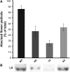
Figure 1.
Figure 1.
Abscission of Flower Pedicels Is Delayed in LX-Inhibited Transgenic Tomato Plants. Abscission of flower pedicels was induced in the wild type (WT) (VF36) and three independent LX-inhibited tomato lines (H9, T2, and A2) by flower removal. (A) The accumulated number of abscising pedicels after 24 h was recorded, and the percentages of abscising pedicels are presented. Four biologically independent treatments were performed in each one of the plant lines, and the total number of pedicels scored was 40 to 60. Error bars correspond to ± se. (B) Level of the LX protein in AZ-containing tissue collected from the different plants 5 h after induction of abscission. LX level was measured following protein extraction by immunoblot analysis.
Figure 2.
Figure 2.
Effects of Flower Removal for Induction of Pedicel Abscission, 1-MCP Pretreatment, and IAA Application after Flower Removal on the Kinetics of Changes in Array-Measured LX Expression Levels during Tomato Pedicel Abscission.
Figure 3.
Figure 3.
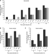
Immunolocalization of LX in Tomato Leaf Petiole AZ, Measurement of LX Level in the Flower Pedicel AZ and NAZ, and Measurement of LX Gene Expression Levels in Flower Pedicel and Leaf Petiole AZs. Longitudinal sections of tomato leaf AZ during the cell separation stage (36 h after ethylene induction), including the stem and leaf petiole tissues, were subjected to immunolocalization of LX using an LX-specific antibody. Immunolocalization of LX in tomato leaf petiole AZ ([A] to [C]), measurement of LX level in the flower pedicel AZ and NAZ (D), and measurement of LX gene expression levels in flower pedicel (E) and leaf petiole (F) AZs. (A) Dark-purple staining indicates cells cross-reacting positively with the LX protein antibody; proximal and distal sides of the AZ fracture plane face the stem and leaf petiole, respectively. (B) Magnification of the AZ tissue in which cell separation has already occurred, where only cells at the edge of the distal side are positively stained with the LX antibody. Bar = 200 μm. (C) Magnification of a sequential serial section of the AZ tissue, in which the immunolocalization assay was performed with preimmune serum showing no LX-specific staining. Bar = 200 μm. (D) Immunoblot analysis using proteins extracted from tissue sampled separately from either the proximal or distal sides of the tomato flower pedicel AZ, and proteins extracted from nearby (a few millimeters away) stem tissue as a control (NAZ). (E) and (F) RNA was extracted from either the proximal or distal sides of the AZ and used for quantitative real-time PCR analysis performed for flower (E) and leaf (F) AZ. Error bars correspond to ±sd. [See online article for color version of this figure.]
Figure 4.
Figure 4.
Changes in Nuclear Morphology in the Tomato Leaf and Flower AZs and Nuclear DNA Fragmentation in Cells on the Distal Side of the AZ. For observing nuclear morphology, abscission was induced either by flower removal or leaf deblading followed by a 24-h exposure to ethylene, and the pedicel or petiole AZs were separated at the late stage of the process. The exposed AZ fracture plane surfaces were subjected to DAPI nuclear staining. (A) Representative images are shown of the fracture plane of the leaf AZ not induced for abscission, including a scaled-up image of one representative nucleus. (B) Image of the leaf AZ at the late stage of induced abscission, including a scaled-up image of one representative nucleus. (C) Image of the flower AZ not induced for abscission. (D) Image of the flower AZ at the late stage of induced abscission. (E) and (F) For detection of DNA fragmentation, a longitudinal section of tomato leaf AZ during the cell separation stage (24 h after ethylene treatment), including the stem and leaf petiole tissues, was stained by TUNEL-labeling assay (E) or by DAPI (F). TUNEL-positive nuclei, characterized by bright-green fluorescence, are localized on the distal side of the leaf AZ (toward the leaf petiole). White dotted lines outline the fracture in the AZ formed due to cell separation.
Figure 5.
Figure 5.
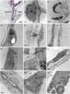
Transmission Electron Micrographs of Cells in Tomato Leaf AZ Showing Ultrastructural Changes 48 h after Induction of Abscission by Leaf Deblading and Ethylene Treatment. The position of cells within the tissue is labeled on the light microscopy image in the top left corner (arrow marks the AZ fracture plane). Asterisks mark detachment of plasma membrane from the cell wall. Cell organelle labeling: c, chloroplast; cw, cell wall; er, endoplasmic reticulum; ga, Golgi apparatus; lv, lytic vacuole; m, mitochondria; n, nucleus; p, plasmodesmata; pb, paramural body; v, vacuole. (A) Intact control taken from the middle of the AZ in untreated plant. (B) to (G) Proximal side of the leaf treated with ethylene. (B) Extensive portion of the ER. (C) Branched plasmodesmata with enlarged cavity connected to ER on both sides of the cell wall. (D) Ameboidal nucleus. (E) Branched plasmodesmata connected to the PB. (F) Branched plasmodesmata with cavities that are not connected to the PB. (G) Cells on the proximal side after releasing the petiole, with retained extensive membrane and vesicular portion. (H) to (J) Distal side of the leaf treated with ethylene. (H) Shrunken chloroplasts and disintegrated cytoplasm. (I) Dilated ER. (J) Condensed nucleus. (K) Cells at the fracture front with swollen chloroplasts containing plastoglobuli. [See online article for color version of this figure.]
Figure 6.
Figure 6.
Nuclease and Protease Activities and Their Encoding Genes Are Induced in the AZ following Induction of Flower and Leaf Abscission. (A) Nuclease activity induced in the tomato flower pedicel and tree tobacco leaf petiole AZs. Total proteins (5 μg) extracted from AZ tissue or nearby NAZ tissue as a control were subjected to nuclease activity gel assay. Arrowhead marks position of the AZ-induced nuclease activity. (B) Nuclease activity induced on the distal side of the tomato flower pedicel AZ. Proteins (5 μg) extracted from either the AZ proximal or distal tissue were used for the nuclease activity gel assay. (C) Quantitative real-time PCR analysis for the nuclease gene TBN1 performed with RNA extracted from tomato tissues sampled from either the proximal or distal sides of the AZ at the indicated times following induction of pedicel abscission by flower removal. (D) Protease activity induced in tomato leaf AZ. Proteins (10 μg), extracted from the AZ or NAZ tissues of mature leaf petiole approaching abscission and young leaf petiole were subjected to protease activity gel assay. Arrowhead marks position of the AZ-induced protease activity. (E) and (F) Microarray analysis results are shown for genes encoding Cys protease (E) and Ser protease (F) in the AZ and NAZ tissues, with or without 1-MCP pretreatment. The results demonstrate induction of expression mainly in the tomato flower pedicel AZ following induction of the pedicel abscission process by flower removal. Error bars correspond to ±sd. [See online article for color version of this figure.]
Figure 7.
Figure 7.
ROS Levels and NADPH Oxidase Gene Expression Are Induced in the Tomato Flower AZ following Induction of Pedicel Abscission by Flower Removal. (A) to (H) ROS staining with DCF was performed on AZ cross sections in control flower AZ not induced for abscission (B) and ~10 h after induction of pedicel abscission by flower removal (D). Bright-field images of the same control (A) and abscission-induced (C) tissue sections. ROS detection was performed also in the exposed AZ surface separating the proximal and distal sides, and fluorescent images of the fracture planes were taken. Representative images of the proximal (E) and distal (G) sides of the pedicel AZ not induced for abscission and of the proximal (F) and distal (H) side of the pedicel AZ induced for abscission are shown. (I) Quantitative real-time PCR expression analysis of the tomato NADPH oxidase gene in the flower AZ during pedicel abscission. Error bars correspond to ±sd. [See online article for color version of this figure.]
Figure 8.
Figure 8.
Organ Abscission Is Delayed in Tomato Transgenic Plants Overexpressing an IAP. Leaf petiole (A) and flower pedicel (B) abscission in two independent transgenic plants overexpressing the IAP gene compared with that measured in the respective Rutgers wild type (WT). Flower pedicel abscission in plants overexpressing the P35 IAP gene compared with that measured in the respective asc wild type (C). Leaf abscission experiments were performed with plants developing six to eight leaves. Petiole abscission of the first leaf was induced by leaf deblading, followed by exposure to ethylene for 24 h. Pedicel abscission was induced by flower removal. Error bars correspond to ±sd.
Figure 9.
Figure 9.
TAPG1, TAPG4, Cel1, and ACS Genes Are Differentially Expressed during Pedicel Abscission in Tissues Localized Distal or Proximal to the Tomato Flower AZ. Tissues were sampled at the indicated time points following flower removal for abscission induction from either the proximal or distal side of the flower AZ, and extracted RNA was used for quantitative real-time PCR. Error bars correspond to ±sd. The accession IDs for the examined genes are as follows: Cel1, U13054; TAPG1, AF001000; TAPG4, U70481; and ACS, M34289. [See online article for color version of this figure.]

Similar articles

Cited by

References

    1. Abeles F.B., Rubinstein B. (1964). Regulation of ethylene evolution and leaf abscission by auxin. Plant Physiol. 39: 963–969 - PMC - PubMed
    1. Agustí J., Merelo P., Cercós M., Tadeo F.R., Talón M. (2009). Comparative transcriptional survey between laser-microdissected cells from laminar abscission zone and petiolar cortical tissue during ethylene-promoted abscission in citrus leaves. BMC Plant Biol. 9: 127. - PMC - PubMed
    1. Aleksandrushkina N.I., Vanyushin B.F. (2009). Endonucleases and their involvement in plant apoptosis. Russ. J. Plant Physiol. 56: 291–305
    1. An Q., Ehlers K., Kogel K.H., van Bel A.J.E., Hückelhoven R. (2006). Multivesicular compartments proliferate in susceptible and resistant MLA12-barley leaves in response to infection by the biotrophic powdery mildew fungus. New Phytol. 172: 563–576 - PubMed
    1. An Q., van Bel A.J., Hückelhoven R. (2007). Do plant cells secrete exosomes derived from multivesicular bodies? Plant Signal. Behav. 2: 4–7 - PMC - PubMed

Publication types

MeSH terms

LinkOut - more resources