

Regional grey and white matter volumetric changes in myalgic encephalomyelitis (chronic fatigue syndrome): a voxel-based morphometry 3 T MRI study
- PMID: 22128128
- PMCID: PMC3474083
- DOI: 10.1259/bjr/93889091
Regional grey and white matter volumetric changes in myalgic encephalomyelitis (chronic fatigue syndrome): a voxel-based morphometry 3 T MRI study
Abstract
Objective: It is not established whether myalgic encephalomyelitis/chronic fatigue syndrome (CFS) is associated with structural brain changes. The aim of this study was to investigate this by conducting the largest voxel-based morphometry study to date in CFS.
Methods: High-resolution structural 3 T cerebral MRI scanning was carried out in 26 patients with CFS and 26 age- and gender-matched healthy volunteers. Voxel-wise generalised linear modelling was applied to the processed MR data using permutation-based non-parametric testing, forming clusters at t>2.3 and testing clusters for significance at p<0.05, corrected for multiple comparisons across space.
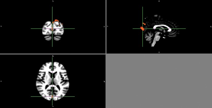
Results: Significant voxels (p<0.05, corrected for multiple comparisons) depicting reduced grey matter volume in the CFS group were noted in the occipital lobes (right and left occipital poles; left lateral occipital cortex, superior division; and left supracalcrine cortex), the right angular gyrus and the posterior division of the left parahippocampal gyrus. Significant voxels (p<0.05, corrected for multiple comparisons) depicting reduced white matter volume in the CFS group were also noted in the left occipital lobe.
Conclusion: These data support the hypothesis that significant neuroanatomical changes occur in CFS, and are consistent with the complaint of impaired memory that is common in this illness; they also suggest that subtle abnormalities in visual processing, and discrepancies between intended actions and consequent movements, may occur in CFS.
Figures
References
-
- Fukuda K, Straus SE, Hickie I, Sharpe MC, Dobbins JG, Komaroff A, et al. The chronic fatigue syndrome: a comprehensive approach to its definition and study. Ann Intern Med 1994;121:953–9 - PubMed
-
- Natelson BH, Cohen JM, Brassloff I, Lee HJ. A controlled study of brain magnetic resonance imaging in patients with the chronic fatigue syndrome. J Neurol Sci 1993;120:213–17 - PubMed
-
- Lange G, DeLuca J, Maldjian JA, Lee H, Tiersky LA, Natelson BH. Brain MRI abnormalities exist in a subset of patients with chronic fatigue syndrome. J Neurol Sci 1999;171:3–7 - PubMed
Publication types
MeSH terms
Grants and funding
LinkOut - more resources
Full Text Sources
Medical
