. 2012 Jan 13;287(3):1915-22.
doi: 10.1074/jbc.M111.279364. Epub 2011 Nov 29.

Interactions between the conserved hydrophobic region of the prion protein and dodecylphosphocholine micelles

Affiliations

Interactions between the conserved hydrophobic region of the prion protein and dodecylphosphocholine micelles

Simon Sauvé et al. J Biol Chem. .

Abstract

The three-dimensional structure of PrP110-136, a peptide encompassing the conserved hydrophobic region of the human prion protein, has been determined at high resolution in dodecylphosphocholine micelles by NMR. The results support the conclusion that the (Ctm)PrP, a transmembrane form of the prion protein, adopts a different conformation than the reported structures of the normal prion protein determined in solution. Paramagnetic relaxation enhancement studies with gadolinium-diethylenetriaminepentaacetic acid indicated that the conserved hydrophobic region peptide is not inserted symmetrically in the micelle, thus suggesting the presence of a guanidium-phosphate ion pair involving the side chain of the terminal arginine and the detergent headgroup. Titration of dodecylphosphocholine into a solution of PrP110-136 revealed the presence of a surface-bound species. In addition, paramagnetic probes located the surface-bound peptide somewhere below the micelle-water interface when using the inserted helix as a positional reference. This localization of the unknown population would allow a similar ion pair interaction.


Figures

FIGURE 1.
FIGURE 1.
Two-dimensional 1H,15N HSQC of uniformly 13C,15N- labeled PrP 110–136 at 600 MHz, 1 mm peptide in 10 mm NaPi, pH 7.6, with 75 mm DPC, recorded at 37 °C. Resonances of the backbone amides are labeled according to the Syrian hamster prion protein sequence.
FIGURE 2.
FIGURE 2.
Solution structure of PrP(110–136) in DPC micelles. A, ensemble of 20 lowest energy conformer calculated using NOE-derived constraints and dihedral angles obtained from TALOS+. B, ribbon representation of the first conformer from the ensemble.
FIGURE 3.
FIGURE 3.
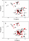
Two-dimensional 1H,15N HSQC spectra recorded after adding the first aliquot of DPC (14 mm) to a 1 mm (A) and a 2 mm (B) sample of PrP(110–136). The black and red contours correspond to the folded peptide and the unknown species, respectively. The folded-to-unknown ratio is ∼ 1:1 in A and ∼1:4 in B. The intensities of the folded resonances appear lower in B because the contour level was lowered for clarity.
FIGURE 4.
FIGURE 4.
Plots of the normalized intensities of peptide resonances as a function of the paramagnetic relaxation agent (PRA) concentration recorded on sample containing 2 mm PrP(110–136) and 2.5 mm DPC. A, Gd-DTPA titration of selected resonances of the folded PrP. B, PRA titration of all well resolved resonances of the unknown conformer. A rapid decrease of signal intensity is indicative of an amide pair in close proximity to the PRA whereas a slow decrease is indicative of an amide pair that is well buried into the DPC micelle. Lines are color coded to reflect the effect of the PRA: blue is least affected (most buried), and red is most affected. The black lines describe the decay of the resonances (not assigned) to the unknown species. The color scheme is applied to the residues of the peptide sequence. Data points are fitted to a single exponential with a y intercept set to 1.0.
FIGURE 5.
FIGURE 5.
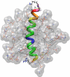
Modeling PrP(110–136) in a micelle of DPC. The average NMR structure of the peptide was manually positioned in a micelle containing 54 DPC molecules obtained by molecular dynamics simulation (see “Results”). The position of the peptide was based on the results of the PRE experiments. The blue residues were positioned at the center of mass of the micelle. This resulted in alignment of the yellow residues with the phosphate group of the detergent headgroup.
FIGURE 6.
FIGURE 6.
Proposed folding of the CHR followed by the binding of the unknown conformer at the micelle surface (see “Discussion”).

Similar articles

Cited by

References

    1. Will R. G., Alpers M. P., Dormont D., Schonberger L. B. (2004) Infectious and Sporadic Prion Diseases in Prion Biology and Diseases (Pruisner S. B., ed) p. 631, Cold Spring Harbor Laboratory, Cold Spring Harbor, NY
    1. Cobb N. J., Surewicz W. K. (2009) Prion diseases and their biochemical mechanisms. Biochemistry 48, 2574–2585 - PMC - PubMed
    1. Hodak M., Chisnell R., Lu W., Bernholc J. (2009) Functional implications of multistage copper binding to the prion protein. Proc. Natl. Acad. Sci. U.S.A. 106, 11576–11581 - PMC - PubMed
    1. Bremer J., Baumann F., Tiberi C., Wessig C., Fischer H., Schwarz P., Steele A. D., Toyka K. V., Nave K. A., Weis J., Aguzzi A. (2010) Axonal prion protein is required for peripheral myelin maintenance. Nat. Neurosci. 13, 310–318 - PubMed
    1. Zahn R., Liu A., Lührs T., Riek R., von Schroetter C., López García F., Billeter M., Calzolai L., Wider G., Wüthrich K. (2000) NMR solution structure of the human prion protein. Proc. Natl. Acad. Sci. U.S.A. 97, 145–150 - PMC - PubMed

Publication types

MeSH terms