. 2012 Jan 13;287(3):1742-54.
doi: 10.1074/jbc.M111.235010. Epub 2011 Nov 29.

Heterogeneous nuclear ribonucleoprotein (hnRNP) F is a novel component of oligodendroglial RNA transport granules contributing to regulation of myelin basic protein (MBP) synthesis

Affiliations

Heterogeneous nuclear ribonucleoprotein (hnRNP) F is a novel component of oligodendroglial RNA transport granules contributing to regulation of myelin basic protein (MBP) synthesis

Robin White et al. J Biol Chem. .

Abstract

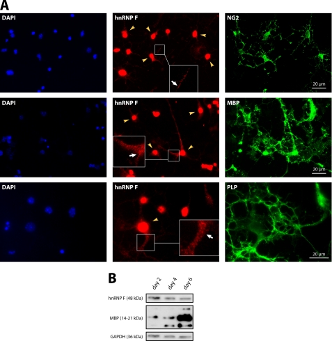
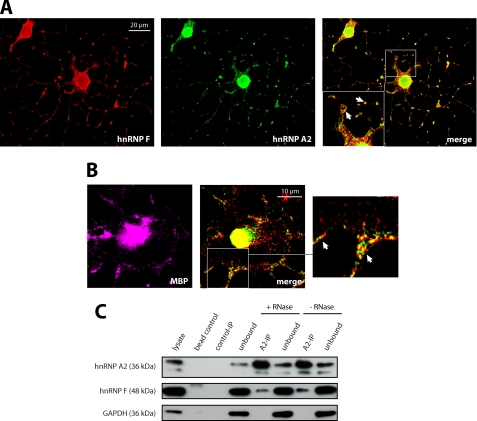
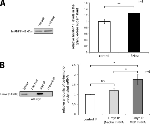
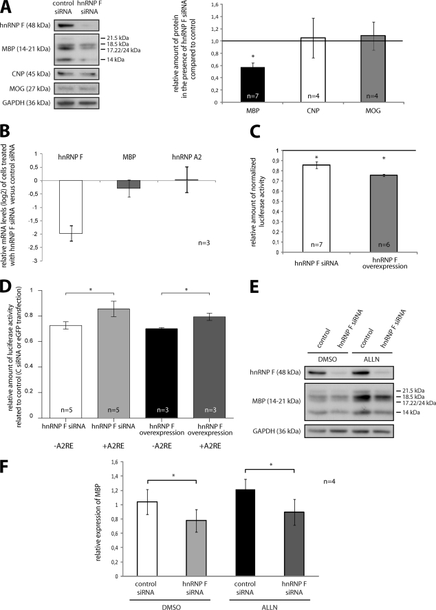
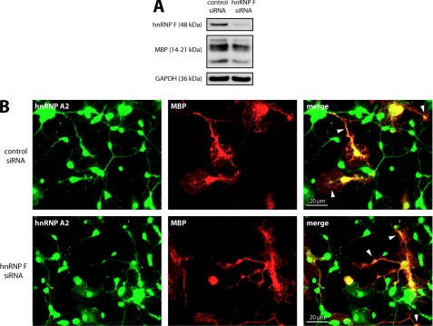
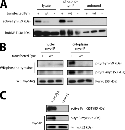
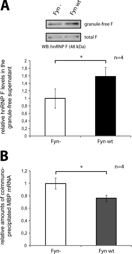
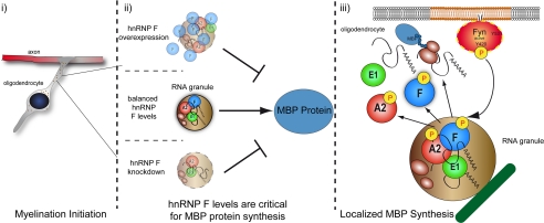
Myelin basic protein (MBP) is a major component of central nervous system (CNS) myelin. The absence of MBP results in the loss of almost all compact myelin in the CNS. MBP mRNA is sorted into RNA granules that are transported to the periphery of oligodendrocytes in a translationally inactive state. A central mediator of this transport process is the trans-acting factor heterogeneous nuclear ribonucleoprotein (hnRNP) A2 that binds to the cis-acting A2-response element in the 3'UTR of MBP mRNA. Recently, we found that activation of the Src family nonreceptor tyrosine kinase Fyn in oligodendrocytes leads to phosphorylation of hnRNP A2 and to increased translation of MBP mRNA. Here, we identify the RNA-binding protein hnRNP F as a novel component of MBP mRNA transport granules. It is associated with hnRNP A2 and MBP mRNA in cytoplasmic granular structures and is involved in post-transcriptional regulation of MBP expression. Fyn kinase activity results in phosphorylation of hnRNP F in the cytoplasm and its release from MBP mRNA and RNA granules. Our results define hnRNP F as a regulatory element of MBP expression in oligodendrocytes and imply an important function of hnRNP F in the control of myelin synthesis.


Figures

FIGURE 1.
FIGURE 1.
hnRNP F is present in the nucleus and the cytoplasm of oligodendrocytes. A, in addition to its prominent nuclear localization (yellow arrowheads), hnRNP F is present in granular structures throughout the cytoplasm (white arrows). Primary mouse oligodendrocytes after 2 or 4 days in culture were stained with antibodies to hnRNP F and markers for oligodendrocyte precursor cells (NG2; 2 days in vitro) or more mature oligodendrocytes (MBP, PLP; 4 days in vitro). Cell nuclei were stained with DAPI. Insets show enlarged areas. B, Western blots confirming the expression of hnRNP F in immature and mature oligodendrocytes (2, 4, and 6 days in vitro). MBP serves as marker for the ongoing differentiation of the cells, and GAPDH is shown as a loading control.
FIGURE 2.
FIGURE 2.
hnRNP F and hnRNP A2 associate in cytoplasmic granular structures in oligodendroglial cells. A, partial colocalization of hnRNP F and hnRNP A2 in granular structures (white arrows) in the cytoplasm of Oli-neu cells that were allowed to differentiate for 3 days and immunostained for hnRNP F and hnRNP A2. Single deconvoluted planes are depicted; the inset shows an enlarged area. B, partial colocalization of hnRNP F and hnRNP A2 in granular structures (enlargement: white arrows) in the cytoplasm of primary mouse oligodendrocytes that were immunostained for hnRNP F and hnRNP A2 after 3 days in culture. MBP is shown as oligodendroglial marker. Single deconvoluted planes are depicted. C, hnRNP F coimmunoprecipitates with hnRNP A2. Immunoprecipitations with hnRNP A2 or isotype-matched control antibodies were performed from RNase- or untreated Oli-neu postnuclear lysates and analyzed on Western blots. The absence of GAPDH in the hnRNP A2-IP confirms the specificity of hnRNP F binding. Note that the hnRNP A2 Western blots show an additional lower band at ∼32 kDa, which is likely to be the hnRNP A2/B1 splice variant hnRNP A2b.
FIGURE 3.
FIGURE 3.
hnRNP F is associated with MBP mRNA and RNA granules. A, disruption of RNA granules increases the amount of soluble cytoplasmic hnRNP F. Postnuclear lysates of Oli-neu cells were treated with RNase A, and after depletion of RNA granules by ultracentrifugation, soluble cytoplasmic proteins were analyzed on Western blots. The change in soluble hnRNP F protein was densitometrically quantified and normalized to GAPDH. **, p < 0.01 (Student's t test). B, MBP mRNA coimmunoprecipitates with hnRNP F-myc. Oli-neu cells were cotransfected with hnRNP F-myc (F-myc) and MBP14 including its 3′UTR. After differentiation for 3 days, immunoprecipitations with antibodies against the Myc tag or isotype-matched control antibodies were performed from postnuclear lysates, and associated mRNAs were analyzed by qRT-PCR. MBP and β-actin mRNA levels were normalized to phosphoglycerate kinase 1 mRNA. n.s., not significant. *, p < 0.05 (Wilcoxon signed-rank test).
FIGURE 4.
FIGURE 4.
MBP synthesis is influenced by hnRNP F. A, knockdown of hnRNP F specifically affects MBP levels. Primary mouse oligodendrocytes were treated with hnRNP F or control siRNA and allowed to differentiate for 3 days. Protein levels were quantified by densitometric analysis of Western blots, and the expression of MBP, CNP, and MOG was normalized to GAPDH. B, knockdown of hnRNP F has no impact on levels of MBP mRNA. Primary mouse oligodendrocytes were treated as in A, but here mRNA levels were analyzed by qRT-PCR and normalized to β-actin mRNA. C, altered hnRNP F levels impair the translation of MBP luciferase reporters. hnRNP F levels in Oli-neu cells were reduced by transfection of hnRNP F-directed siRNA or increased by transfection of hnRNP F expression vectors. Subsequently, luciferase-based translational reporters containing parts of the 3′UTR of MBP as regulatory elements were used to measure translational activity in DualGlo assays (see “Experimental Procedures” for details). Relative luciferase activity is reduced in cells treated with hnRNP F siRNA as well as in hnRNP F-overexpressing cells that were related to control siRNA- or GFP-transfected cells, respectively. D, effect of hnRNP F levels on the translation of MBP reporters, including or lacking the A2RE. Cells were treated as in C, but here a luciferase reporter construct lacking the A2RE was included. E, reduced MBP levels in response to hnRNP F knockdown are independent of proteasomal activity in primary oligodendrocytes. The experiment was performed according to A. Before lysis, the siRNA-treated cells were incubated with the proteasomal inhibitor ALLN or as control with DMSO. F, statistical evaluation of four independent experiments as shown in E. Note the persistence in MBP reduction despite inhibition of the proteasome. *, p < 0.05 (Student's t test (D and F) or Wilcoxon signed-rank test (A and C)).
FIGURE 5.
FIGURE 5.
Cytoplasmic localization of hnRNP A2 and MBP distribution appear unaffected by hnRNP F knockdown. A, primary mouse oligodendrocytes were treated with hnRNP F or control siRNA and allowed to differentiate for 3 days. The knockdown of hnRNP F was assessed by Western analysis. GAPDH serves as loading control. B, cells from A were immunostained for hnRNP A2 and MBP. Note the characteristic distribution of MBP and the cytoplasmic localization of hnRNP A2, in particular its concentration at foci where MBP is highly abundant (white arrowheads).
FIGURE 6.
FIGURE 6.
Cytoplasmic hnRNP F is a target of Fyn kinase. A, hnRNP F is tyrosine-phosphorylated in response to Fyn activity. Oli-neu cells were transfected with constitutive active (Fyn+), wild type (Fyn WT), or kinase-inactive (Fyn) Fyn constructs. After 2 days, tyrosine-phosphorylated proteins were immunoprecipitated and analyzed on Western blots together with total lysates and proteins that were not precipitated (unbound). B, Fyn-dependent tyrosine phosphorylation of hnRNP F occurs in the cytoplasm of oligodendrocytes. Oli-neu cells were cotransfected with Fyn+, Fyn WT, or Fyn together with Myc-tagged hnRNP F (F-myc). 2 days later, separate nuclear and cytoplasmic fractions were prepared, and immunoprecipitations with antibodies against the Myc tag were performed. The immunoprecipitations were analyzed on Western blots for tyrosine-phosphorylated proteins and total amounts of F-myc. C, Fyn phosphorylates hnRNP F directly. Myc-tagged hnRNP F was immunoprecipitated and incubated in the presence or absence of recombinant Fyn. Western blot analysis with phosphotyrosine- and Myc-specific antibodies shows that the addition of Fyn results in tyrosine phosphorylation of hnRNP F. An activity-dependent antibody (Src-pY418) demonstrates that recombinant Fyn is active in the experiment.
FIGURE 7.
FIGURE 7.
Fyn activity leads to a release of hnRNP F from MBP mRNA. A, Fyn activity releases hnRNP F from the granule fraction into the cytosol. Oli-neu cells were transfected with Fyn WT or Fyn constructs. After 2 days, postnuclear total lysates and granule-free lysates, obtained by ultracentrifugation, were analyzed on Western blots. The levels of hnRNP F in the nongranule fraction were quantified densitometrically and normalized to total hnRNP F levels. B, Fyn activity releases hnRNP F from MBP mRNA. Oli-neu cells were cotransfected with F-myc and Fyn WT or Fyn, respectively. After differentiation for 3 days, immunoprecipitations with antibodies against the Myc tag or control antibodies were performed from postnuclear lysates, and associated mRNAs were analyzed by qRT-PCR. MBP mRNA levels were normalized to levels of phosphoglycerate kinase 1 mRNA. *, p < 0.05 (Student's t test).
FIGURE 8.
FIGURE 8.
Model. MBP mRNA is transported in RNA granules toward the periphery of the cell where Fyn-mediated translational initiation occurs by phosphorylation of RNA-binding proteins and their liberation from the granule and a dissociation of MBP mRNA. Distinct levels of hnRNP F seem to be required to form fully functional MBP mRNA granules thus facilitating efficient protein synthesis.

Similar articles

Cited by

References

    1. Nave K. A. (2010) Myelination and the trophic support of long axons. Nat. Rev. Neurosci 11, 275–283 - PubMed
    1. Aggarwal S., Yurlova L., Simons M. (2011) Central nervous system myelin. Structure, synthesis, and assembly. Trends Cell Biol. 21, 585–593 - PubMed
    1. Emery B. (2010) Regulation of oligodendrocyte differentiation and myelination. Science 330, 779–782 - PubMed
    1. Simons M., Trotter J. (2007) Wrapping it up. The cell biology of myelination. Curr. Opin. Neurobiol. 17, 533–540 - PubMed
    1. Griffiths I., Klugmann M., Anderson T., Yool D., Thomson C., Schwab M. H., Schneider A., Zimmermann F., McCulloch M., Nadon N., Nave K. A. (1998) Axonal swellings and degeneration in mice lacking the major proteolipid of myelin. Science 280, 1610–1613 - PubMed

Publication types

MeSH terms

Substances

LinkOut - more resources