. 2012 Jan 13;287(3):1734-41.
doi: 10.1074/jbc.M111.308650. Epub 2011 Nov 28.

Amer2 protein is a novel negative regulator of Wnt/β-catenin signaling involved in neuroectodermal patterning

Affiliations

Amer2 protein is a novel negative regulator of Wnt/β-catenin signaling involved in neuroectodermal patterning

Astrid S Pfister et al. J Biol Chem. .

Abstract

Wnt/β-catenin signaling is negatively controlled by the adenomatous polyposis coli (APC) tumor suppressor, which induces proteasomal degradation of β-catenin as part of the β-catenin destruction complex. Amer2 (APC membrane recruitment 2; FAM123A) is a direct interaction partner of APC, related to the tumor suppressor Amer1/WTX, but its function in Wnt signaling is not known. Here, we show that Amer2 recruits APC to the plasma membrane by binding to phosphatidylinositol 4,5-bisphosphate lipids via lysine-rich motifs and that APC links β-catenin and the destruction complex components axin and conductin to Amer2. Knockdown of Amer2 increased Wnt target gene expression and reporter activity in cell lines, and overexpression reduced reporter activity, which required membrane association of Amer2. In Xenopus embryos, Amer2 is expressed mainly in the dorsal neuroectoderm and neural tissues. Down-regulation of Amer2 by specific morpholino oligonucleotides altered neuroectodermal patterning, which could be rescued by expression of a dominant-negative mutant of Lef1 that interferes with β-catenin-dependent transcription. Our data characterize Amer2 for the first time as a negative regulator of Wnt signaling both in cell lines and in vivo and define Amer proteins as a novel family of Wnt pathway regulators.


Figures

FIGURE 1.
FIGURE 1.
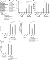
Amer2 interacts with β-catenin destruction complex, localizes to plasma membrane, and binds to phosphatidylinositol 4,5-bisphosphate. A, schematic representation of the Amer2 protein. APC-interacting domains A1 and A2 are shown in gray. K1 and K2 denote two clusters of highly conserved lysine residues indicated by black lines (cf. supplemental Fig. S3). Amino acids are depicted. B, FLAG-tagged Amer2 interacted with EGFP-APC-Arm but not with mutant EGFP-APC-Arm-N507K in co-immunoprecipitation experiments after transient transfection of HEK293T cells. Immunoprecipitation (IP) was performed using FLAG-Sepharose beads, and Western blots were detected using mouse anti-GFP and rabbit anti-FLAG antibodies. Amer1 served as a control. The asterisk indicates the two splice variants of Amer2 generated form the cDNA (cf. supplemental Fig. S2). C, Amer2 recruited APC to the plasma membrane, which required an APC-binding domain. Fluorescence micrographs are shown of MCF-7 cells transiently transfected with EGFP-tagged Amer2(2–421) or Amer2(2–315) and CMV-APC as indicated. Cells were stained with anti-APC serum, and Amer2 was detected by fluorescence. The arrowheads point to the colocalization of Amer2 and APC at the plasma membrane. D, β-catenin, axin, and conductin co-immunoprecipitated with Amer2 in the presence (but not absence) of APC after transient transfection in HEK293T cells as indicated. Immunoprecipitation was performed using anti-FLAG beads, and Western blotting was performed using anti-GFP and anti-FLAG antibodies. Arrowheads indicate the positions of β-catenin (β), axin (A), and conductin (C). E, fluorescence micrographs of EGFP-tagged Amer2 mutants as indicated. F, Amer2(2–230) interacted with phosphoinositides. Membrane lipid strips were incubated with recombinantly expressed GST-Amer2(2–230) or GST as a control and detected with anti-GST antibodies. PtdIns, phosphatidylinositol; DAG, diacylglycerol; PA, phosphatidic acid; PS, phosphatidylserine; PE, phosphatidylethanolamine, PC, phosphatidylcholine; PG, phosphatidylglycerol.
FIGURE 2.
FIGURE 2.
Amer2 negatively regulates Wnt/β-catenin signaling at the plasma membrane. A and B, mRNA expression of the Wnt target genes conductin (A) and LGR5 (B). RT-PCR was performed with HEK293T cells transiently transfected with control siRNA (GFP siRNA (siGFP)) and siRNA sequences targeting Amer2 for degradation (Amer2-1 (siAmer2-1) and Amer2-2 (siAmer2-2) siRNAs). GAPDH RT-PCR was used for normalization. Numbers between the panels represent the levels of conductin, Amer2 and LGR5 normalized to GAPDH as determined by densitometry. C, Wnt3A treatment or siRNA-mediated knockdown of Amer2 increased β-catenin levels in cytosolic fractions of HEK293T cells as determined by Western blotting. α-Tubulin staining served as a normalization control. Numbers between the panels represent the levels of β-catenin normalized to α-tubulin as determined by densitometry. D, siRNA-mediated knockdown of Amer2 resulted in up-regulation of β-catenin-dependent transcription. HEK293T cells stably expressing a β-catenin-responsive firefly luciferase reporter were transfected with GFP siRNA and Amer2-1 or Amer2-2 siRNA and stimulated with Wnt3A-conditioned medium (Wnt3A-CM) for 4 h. -Fold stimulation of luciferase activity is shown. E, experiment performed as described for D with RKO cells stably expressing the reporter. F and G, overexpression of Amer2 down-regulated the β-catenin-dependent reporter in HEK293T cells upon stimulation with Wnt3A (F) or cotransfection of FLAG-dominant-active LRP6 (daLRP6; G), which was abolished in the Amer2ΔK1,K2 mutant, which was unable to bind to the plasma membrane. Western blots for Amer2 and dominant-active LRP6 from a representative experiment are shown below the graphs. H, overexpression of Amer2 did not down-regulate the β-catenin-dependent reporter in HEK293T cells stimulated by cotransfection of stabilized β-catenin S33Y. The Western blot shows expression of β-catenin S33Y from a representative experiment. In D–H, error bars indicate S.E. Statistical analysis was done using Student's unpaired t test. Statistically significant differences are indicated: *, p < 0.05; **, p < 0.005. n.s., non-significant difference; n, number of independent experiments analyzed.
FIGURE 3.
FIGURE 3.
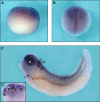
Expression pattern of xAmer2 in Xenopus embryos by whole-mount in situ hybridization. A, xAmer2 expression in Xenopus embryos at NF stage 10.5 in the animal hemisphere (lateral view, animal pole to the top). B, xAmer2 expression in the anterior neural plate at NF stage 16 (dorsal view, anterior to the top). C, xAmer2 expression in the central nervous system, otic vesicle (o), eye (e), and cranial ganglia and nerves (arrowheads; V, trigeminal; VII, facial; IX, glossopharyngeal; X, vagal) at NF stage 35 (lateral view, anterior to the left). C′, cross-section through the head region of an NF stage 35 embryo (anterior to the left). xAmer2 expression was clearly seen in the forebrain (fb), midbrain (mb), and hindbrain (hb) but not the boundaries between these domains (arrowheads).
FIGURE 4.
FIGURE 4.
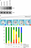
xAmer2 negatively regulates Wnt signaling in vivo. A, depletion of xAmer2 by xAmer2 MO (cf. supplemental Fig. 4A) increased expression of the Wnt target gene Xnr3. Xenopus embryos were injected in both dorsal blastomeres at the four-cell stage with 0.4 pmol of xAmer2 MO or control MO or with 100 pg of β-catenin RNA as a positive control, and Xnr3 mRNA levels were determined by RT-PCR at NF stage 10.5. Ornithine decarboxylase (ODC) RT-PCR was used for normalization. Numbers between the panels represent the levels of Xnr3 normalized to ornithine decarboxylase as determined by densitometry. B, depletion of xAmer2 by xAmer2 MO affected anterior-posterior patterning of the neuroectoderm, consistent with a negative function in Wnt signaling. Embryos were injected in one anterior-dorsal blastomere at the eight-cell stage with 100 pg of lacZ DNA plus 0.2 or 0.4 pmol of Amer2 MO or control MO without or with 500 pg of LefΔBD RNA as indicated. Embryos were stained for lacZ to label the injected side, probed by in situ hybridization for expression of Rx1 and En-2 at NF stage 20, and scored for reduced or expanded expression of the markers on the injected side compared with the uninjected side of the same embryo. Images show embryos representative of the observed phenotypes. Injected sides are oriented to the right in all images. The colored insets represent phenotypes as follows: blue, enhanced Rx1 and En-2; green, equal/normal expression of markers; yellow, markers reduced/anteriorly shifted at the injected side; orange, markers lost at the injected side; red, markers lost at both the injected and uninjected sides of the embryo. The graph shows the statistics of at least three independent experiments. Numbers below the graph represent analyzed embryos.

Similar articles

Cited by

References

    1. MacDonald B. T., Tamai K., He X. (2009) Wnt/β-catenin signaling: components, mechanisms, and diseases. Dev. Cell 17, 9–26 - PMC - PubMed
    1. Polakis P. (2000) Wnt signaling and cancer. Genes Dev. 14, 1837–1851 - PubMed
    1. Grohmann A., Tanneberger K., Alzner A., Schneikert J., Behrens J. (2007) AMER1 regulates the distribution of the tumor suppressor APC between microtubules and the plasma membrane. J. Cell Sci. 120, 3738–3747 - PubMed
    1. Major M. B., Camp N. D., Berndt J. D., Yi X., Goldenberg S. J., Hubbert C., Biechele T. L., Gingras A. C., Zheng N., Maccoss M. J., Angers S., Moon R. T. (2007) Wilms tumor suppressor WTX negatively regulates WNT/β-catenin signaling. Science 316, 1043–1046 - PubMed
    1. Tanneberger K., Pfister A. S., Kriz V., Bryja V., Schambony A., Behrens J. (2011) Structural and functional characterization of the Wnt inhibitor APC membrane recruitment 1 (Amer1). J. Biol. Chem. 286, 19204–19214 - PMC - PubMed

Publication types

MeSH terms