. 2012 Jan 20;287(4):2568-78.
doi: 10.1074/jbc.M111.318766. Epub 2011 Nov 28.

Kinetic analysis of ribosome-bound fluorescent proteins reveals an early, stable, cotranslational folding intermediate

Affiliations

Kinetic analysis of ribosome-bound fluorescent proteins reveals an early, stable, cotranslational folding intermediate

Devaki A Kelkar et al. J Biol Chem. .

Abstract

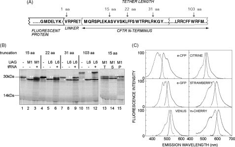
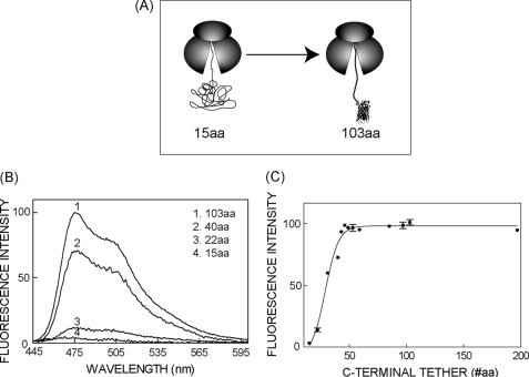
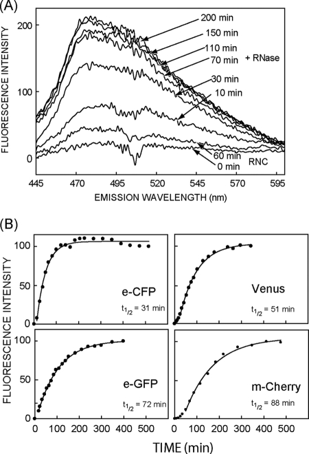
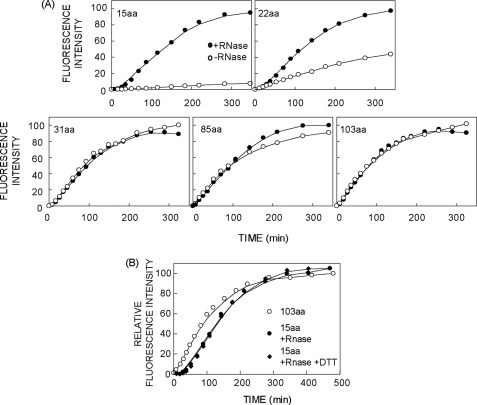
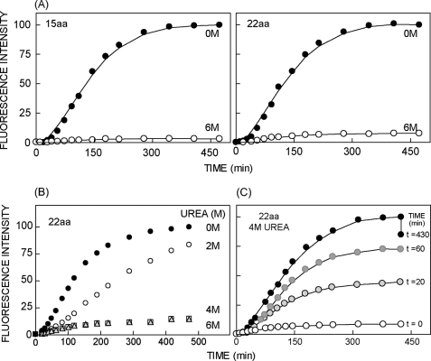
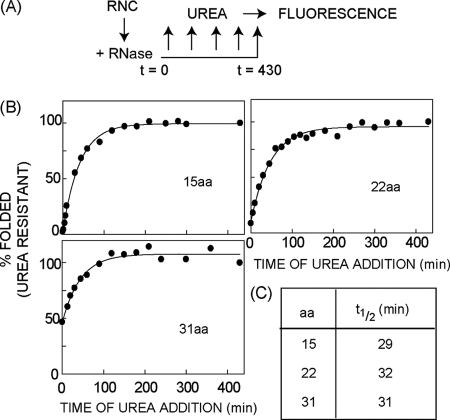
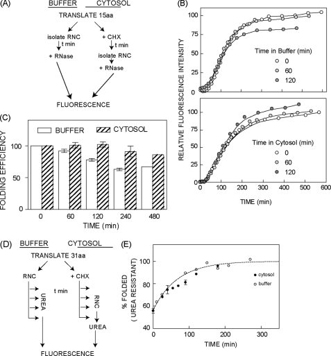
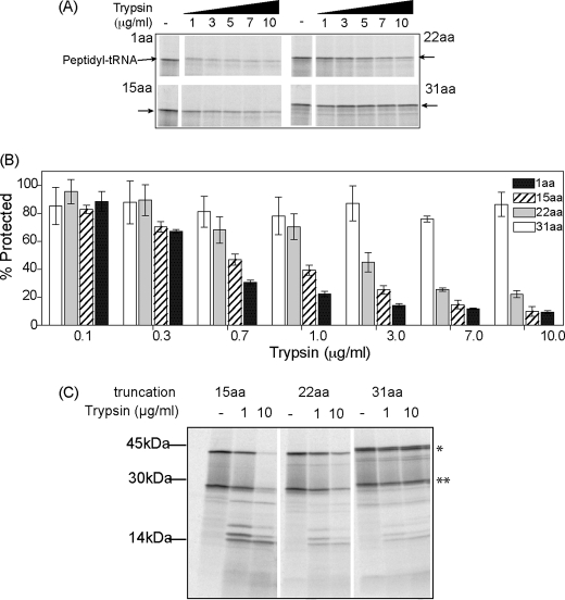
Protein folding in cells reflects a delicate interplay between biophysical properties of the nascent polypeptide, the vectorial nature and rate of translation, molecular crowding, and cellular biosynthetic machinery. To better understand how this complex environment affects de novo folding pathways as they occur in the cell, we expressed β-barrel fluorescent proteins derived from GFP and RFP in an in vitro system that allows direct analysis of cotranslational folding intermediates. Quantitative analysis of ribosome-bound eCFP and mCherry fusion proteins revealed that productive folding exhibits a sharp threshold as the length of polypeptide from the C terminus to the ribosome peptidyltransferase center is increased. Fluorescence spectroscopy, urea denaturation, and limited protease digestion confirmed that sequestration of only 10-15 C-terminal residues within the ribosome exit tunnel effectively prevents stable barrel formation, whereas folding occurs unimpeded when the C terminus is extended beyond the ribosome exit site. Nascent FPs with 10 of the 11 β-strands outside the ribosome exit tunnel acquire a non-native conformation that is remarkably stable in diverse environments. Upon ribosome release, these structural intermediates fold efficiently with kinetics that are unaffected by the cytosolic crowding or cellular chaperones. Our results indicate that during synthesis, fluorescent protein folding is initiated cotranslationally via rapid formation of a highly stable, on-pathway structural intermediate and that the rate-limiting step of folding involves autonomous incorporation of the 11th β-strand into the mature barrel structure.


Figures

FIGURE 1.
FIGURE 1.
In vitro generation and fluorescence of FP RNCs. A, diagram of the FP fusion protein showing the C terminus of the FP, 6-aa linker, and N-terminal residues of CFTR. Truncations sites (arrows) are indicated. B, autoradiogram of 35S-labeled eCFP-CFTR translation products (left panel) with and without UAG codon expressed in the presence or absence of [14C]Lys-tRNAamb (tRNA). Truncation sites are indicated at the top. Polypeptides that terminate or readthrough the UAG codon are indicated by single and double asterisks, respectively. In the right panel, RNCs from the translation (T) were pelleted into the supernatant (S) and pellet (P) fractions. Polypeptides that terminate at UAG (*) are released from the ribosome, whereas readthrough products (**) remain ribosome bound. C, fluorescence excitation and emission spectra obtained from purified FP-RNC complexes containing a 103 C-terminal tether. Spectra were corrected for background (mock translation) and measured >4 h at 24 °C post-purification and arbitrarily normalized to 100.
FIGURE 2.
FIGURE 2.
Nascent FP maturation is constrained by the ribosome exit tunnel. A, diagram of RNCs truncated 15 and 103 aa C-terminal to CFP showing possible conformational states of ribosome attached nascent chains. B, eCFP fluorescence emission spectra obtained from isolated RNCs containing C-terminal tethers of indicated lengths. Spectral intensity was corrected for background signal, and normalized for nascent chain concentration as determined by [14C]Lys incorporation. C, background-corrected peak fluorescence intensity of eCFP-RNCs normalized to chain concentration and plotted as a function of C-terminal tether length. All fluorescence measurements were taken at 4 °C. Where present, error bars represent S.E., n ≥ 3.
FIGURE 3.
FIGURE 3.
De novo FP maturation kinetics following ribosome release. A, eCFP fluorescence emission spectra of isolated RNCs (15 aa tether) prior to (RNC) and after ribosome release by RNase digestion (+RNase). Spectra were measured at 24 °C at the times indicated. B, time-dependent increase in peak fluorescence intensity of eCFP, eGFP, Venus, and mCherry following ribosome release (15 aa tether) at t = 0. Fluorescence at each time point was determined after background correction and normalization to the maximum intensity was obtained. Data were fitted to single exponential kinetics after ignoring the initial “lag” phase. Representative data sets are shown, whereas t½ is the average of at least two independent measurements.
FIGURE 4.
FIGURE 4.
C-terminal tether length constrains mCherry chromophore maturation. A, peak corrected mCherry fluorescence emission intensity was measured at 24 °C at the times indicated using intact RNCs (open circles) and after release of the nascent chain with RNase (filled circles). C-terminal tether length is indicated in each panel. Data were obtained simultaneously for parallel translation samples in each panel. Peak fluorescence intensities were corrected for [14C]Lys content and normalized to the maximum value obtained in each experiment. Spline lines are shown merely as guides to the eye. B, maturation time course of mCherry RNCs measured on intact RNCs containing a 103-aa tether or following ribosome release with RNase with a 15-aa tether in the presence or absence of 0.1 mm DTT. In all experiments, fluorescence intensity was corrected for nascent chain concentration based on [14C]Lys incorporation. Spectra were taken at 24 °C.
FIGURE 5.
FIGURE 5.
Nascent mCherry is sensitive to urea denaturation prior to folding. A, time course of mCherry chromophore maturation in the presence (open circles) or absence (filled circles) of 6 m urea following RNase release of nascent chains (15 and 22 aa tethers). B, mCherry maturation is inhibited in the presence of 4 and 6 m but not 2 m urea. C, mCherry nascent chains (22 aa tether) were released from the ribosome at t = 0 and 4 m urea was added at t = 0, 20, 60, or 430 min. Peak fluorescence intensity at each time point following ribosome release was corrected for nascent chain concentration and normalized to the fluorescence intensity obtained at the last time point in the absence of urea.
FIGURE 6.
FIGURE 6.
De novo folding kinetics of the mCherry β-barrel following ribosome release. A, experimental design used to measure FP folding kinetics as described in Fig. 5. B, β-barrel folding curves based on the acquisition of resistance to 4 m urea. Plot was fitted to single exponential kinetics as described under “Experimental Procedures.” Representative data are shown in B and t½ values in C are averages of at least two independent experiments.
FIGURE 7.
FIGURE 7.
Cytosolic crowding and chaperone machinery do not affect kinetics or stability of nascent mCherry folding intermediates. A, experimental design to measure stability of the nascent mCherry polypeptide. B, maturation time course of mCherry (15 aa tether) following ribosome release after incubation of intact RNCs in buffer (top panel) or RRL translation reaction (bottom panel). Time points refer to the length of incubation prior to ribosome release (buffer) or RNC purification from RRL (cytosol) followed by ribosome release. Background-subtracted peak fluorescence intensity was corrected for [14C]Lys incorporation and normalized to the maximal fluorescence obtained without incubation. C, quantification of folding efficiency determined from experiments in panel B shown normalized to the value obtained at t = 0 incubation time. Data show average of three experiments ± S.E. or 2 experiments (480 min). D, experimental design to measure cytosol effects on folding kinetics. E, folding kinetics of ribosome bound mCherry (31 aa tether) in buffer (open circles) and RRL translation reaction (filled circles). x-Axis refers either to the time of urea addition after RNC isolation in buffer or the duration of RNC incubation in RRL prior to RNC isolation. In each case, fluorescence was measured after maximal chromophore maturation had occurred as described in the legend to Fig. 5. Data shows average of at least two independent experiments and where indicated error bars represent S.E., n ≥ 3.
FIGURE 8.
FIGURE 8.
Conformational sensitivity of kinetically trapped folding intermediates. A, [35S]Met-labeled mCherry translation products were subjected to limited trypsin digestion in RRL. Protease-resistant peptidyl-tRNA bands (arrows) with tether lengths of 1, 15, 22, and 31 aa are shown. B, quantification of trypsin-resistant fragments as shown on panel A was determined by phosphorimaging and expressed as % of the peptidyl-tRNA band present prior to digestion. Data show average of at least three independent experiments ± S.E. C, RNCs were isolated by pelleting and subjected to trypsin digestion as in panel A. In this case, all bands are derived from peptidyl-tRNA, some of which were hydrolyzed during SDS-PAGE and give rise to proteolytic fragments most prominent for nascent chains containing a 15-aa tether.

Similar articles

Cited by

References

    1. Leopold P. E., Montal M., Onuchic J. N. (1992) Protein folding funnels. A kinetic approach to the sequence-structure relationship. Proc. Natl. Acad. Sci. U.S.A. 89, 8721–8725 - PMC - PubMed
    1. Ozkan S. B., Dill K. A., Bahar I. (2002) Fast-folding protein kinetics, hidden intermediates, and the sequential stabilization model. Protein Sci. 11, 1958–1970 - PMC - PubMed
    1. Clark P. L. (2004) Protein folding in the cell. Reshaping the folding funnel. Trends Biochem. Sci. 29, 527–534 - PubMed
    1. Hartl F. U., Hayer-Hartl M. (2009) Converging concepts of protein folding in vitro and in vivo. Nat. Struct. Mol. Biol. 16, 574–581 - PubMed
    1. Kramer G., Boehringer D., Ban N., Bukau B. (2009) The ribosome as a platform for cotranslational processing, folding and targeting of newly synthesized proteins. Nat. Struct. Mol. Biol. 16, 589–597 - PubMed

Publication types

MeSH terms

Substances

LinkOut - more resources