. 2012 Jan 6;287(2):1306-21.
doi: 10.1074/jbc.M111.276865. Epub 2011 Nov 28.

Plasmodium falciparum inhibitor-3 homolog increases protein phosphatase type 1 activity and is essential for parasitic survival

Affiliations

Plasmodium falciparum inhibitor-3 homolog increases protein phosphatase type 1 activity and is essential for parasitic survival

Aline Fréville et al. J Biol Chem. .

Abstract

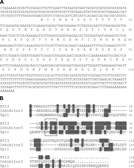
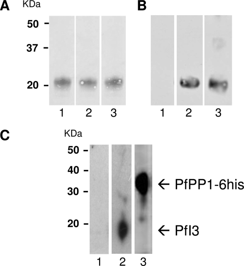
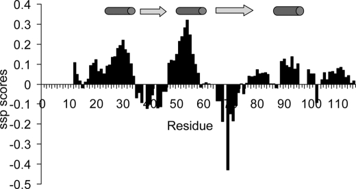
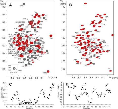
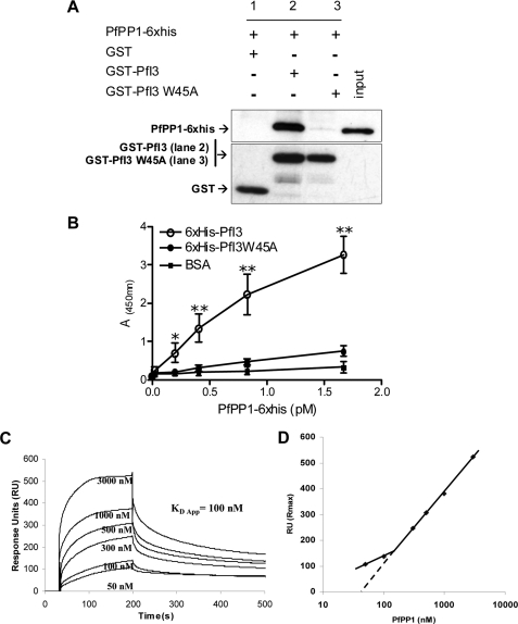
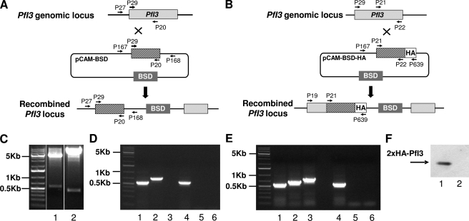
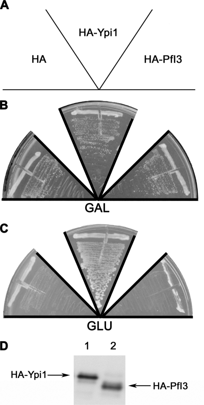
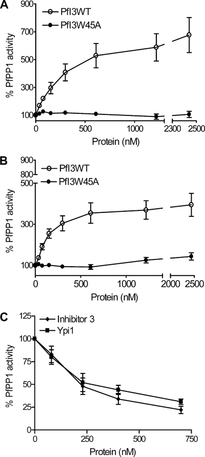
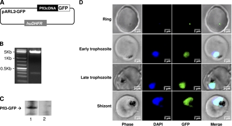
Growing evidence indicates that the protein regulators governing protein phosphatase 1 (PP1) activity have crucial functions because their deletion drastically affects cell growth and division. PP1 has been found to be essential in Plasmodium falciparum, but little is known about its regulators. In this study, we have identified a homolog of Inhibitor-3 of PP1, named PfI3. NMR analysis shows that PfI3 belongs to the disordered protein family. High affinity interaction of PfI3 and PfPP1 is demonstrated in vitro using several methods, with an apparent dissociation constant K(D) of 100 nm. We further show that the conserved (41)KVVRW(45) motif is crucial for this interaction as the replacement of the Trp(45) by an Ala(45) severely decreases the binding to PfPP1. Surprisingly, PfI3 was unable to rescue a yeast strain deficient in I3 (Ypi1). This lack of functional orthology was supported as functional assays in vitro have revealed that PfI3, unlike yeast I3 and human I3, increases PfPP1 activity. Reverse genetic approaches suggest an essential role of PfI3 in the growth and/or survival of blood stage parasites because attempts to obtain knock-out parasites were unsuccessful, although the locus of PfI3 is accessible. The main localization of a GFP-tagged PfI3 in the nucleus of all blood stage parasites is compatible with a regulatory role of PfI3 on the activity of nuclear PfPP1.


Figures

FIGURE 1.
FIGURE 1.
Nucleotide and deduced amino acid sequences of PfI3. A, amino acids are numbered to the right of the sequence. The sequences of 5′- and 3′-untranslated regions were obtained using primers derived from genomic DNA followed by PCR on cDNA and by performing 3′-rapid amplification of cDNA ends, respectively. B, analysis of amino acid sequence of PfI3. PfI3 was aligned with the mammalian (Inhibitor-3) and yeast I3 (Ypi1) homologs using the Megalign program (DNASTAR). The identical residues are shown in gray. The box contains the KVVRW sequence which fits with the consensus sequence (K/R)X0–1(V/I)p(F/W), known as the RVXF motif and required for binding with PP1.
FIGURE 2.
FIGURE 2.
Expression of the PfI3 gene product by P. falciparum. A, purified His fusion PfI3 separated by 15% SDS-PAGE and blotted onto nitrocellulose (Red Ponceau staining, lanes 1–3). B, immunoblot analysis of recombinant PfI3 with rat prebleed sera (lane 1), with rat anti-PfI3 antisera (lane 2), and with mAb anti-His (lane 3) showed a single band at ∼20 Da, indicating an anomalous electrophoretic migration of PfI3 (expected size 13 kDa). The identity of the purified recombinant PfI3 has been further confirmed by MALDI-TOF mass spectrometry. C, detection of endogenous PfI3 in total proteins extracted from asynchronous cultures of P. falciparum. Total protein extracts (10 mg) pre-cleared on Ni-NTA-Sepharose beads were incubated overnight with His6-tagged PfPP1 affinity Ni-NTA column. After washings, proteins eluted with SDS-PAGE loading buffer were migrated and blotted to nitrocellulose. The blots were probed with preimmune serum (lane 1), anti-PfI3 (lane 2), or with anti-His mAb antibodies (lane 3). The blots were revealed as described under “Experimental Procedures.”
FIGURE 3.
FIGURE 3.
Secondary structure propensity scores reported along the PfI3 sequence. Positive values exceeding 0.1 are indicative of transient helices and negative values of extended transient conformations as represented by cylinders and arrows above the graphics, respectively.
FIGURE 4.
FIGURE 4.
NMR mapping of the PfI3 sites interacting with PfPP1. A, overlaid {1H-15N} HSQC of 15N-PfI3, free (gray spectrum), and in presence of an equimolar amount of PfPP1 (superimposed red spectrum). Interaction induces broadening of numerous resonances; see for example the isolated Gly73 resonance, in the upper part of the spectrum. The amide side chain resonance of Trp45 is shown as an inset. B, same color code is applied for the overlaid spectra corresponding to 15N-PfI3W45A free or in presence of PfPP1. Resonances of PfI3 are all annotated (A), although the annotated resonances of 15N-PfI3W45A are restricted to those showing perturbation upon PfPP1 addition or located around Ala45 (B). The supplemental Fig. S2 provides a comparison of the (1H-15N} HSQC of 15N-PfI3 and 15N-PfI3W45A. The graphics below the spectra show the normalized ratio of the intensity of a given resonance in the free PfI3 and in the 1:1 PfI3:PfPP1 spectra (C) or free PfI3W45A and in the 1:1 PfI3W45A:PfPP1 spectra (D), reported along the PfI3 sequence.
FIGURE 5.
FIGURE 5.
Interaction studies of PfI3 with PfPP1 in vitro. A, GST-pulldown assays. Glutathione-agarose beads coupled with GST alone (lane 1), GST-PfI3 bound to beads (lane 2), or GST-PfI3W45A bound beads (lane 3) were incubated with His6-tagged PfPP1. After washes, proteins bound to the beads were separated by 15% SDS-PAGE and blotted to nitrocellulose. Immunoblot analysis was performed with anti-His mAb (upper blot) and mAb anti-GST antibodies (lower blot) providing loading controls for bound GST and GST-fusion proteins. B, quantification of the binding capacity of PfPP1 to PfI3 using an ELISA-based technique. Increased quantities of biotinylated PfPP1 were added to wells coated with recombinant PfI3 or PfI3W45A proteins (1 μg/well). Results represent experiments carried out with two different batches. Bars indicate S.E. (n = 3). *, p < 0.05; **, p < 0.001 when compared with either BSA or PfI3W45A. C, surface plasmon resonance measurements. PfI3 was immobilized on sensor chip, and increasing concentrations of PfPP1 (50–3000 nm) were used for injections. Base line of each sensogram was normalized and expressed in relative RU. Results shown are representative of three experiments. D, plot of the maximum RU reached in the experiment at each analyte concentration (log scale) shows a biphasic nature that can be explained by the presence of several binding sites of different affinities.
FIGURE 6.
FIGURE 6.
Targeted gene disruption and HA tagging of the PfI3 locus. A, gene-targeting construct for gene disruption by single homologous recombination using the pCAM-BSD, and the locus resulting from integration of the knock-out construct. B, epitope tagging of PfI3 by knock-in strategy. Insertion of an HA epitope tag at the C terminus of PfI3 by single homologous recombination (knock-in). The locations of the primers (P29, P20, P21, P22, P27, P167, P168, and P639) used for PCR analysis are indicated as well as the blasticidin-resistance cassette (BSD). C, plasmid rescue experiments showing the presence of pCAM-PfI3 (lane 1) and pCAM-PfI3–2HA (lane 2) constructs in transfected parasite culture. D, analysis of pCAM-PfI3-transfected 3D7 culture by PCR; lanes 1–3 correspond to DNA extracted from transfected parasites; lanes 4–6 correspond to DNA extracted from wild type parasites. Lanes 1 and 4 represent the detection of a portion of the wild type locus (PCR with P29 and P20); lanes 2 and 5 represent the detection of episomal DNA (PCR with P167 and P168); and lanes 3 and 6 represent the detection of the integration at the 5′ end of the insert (PCR with P27 and P168). The absence of amplification of a PCR product using genomic DNA prepared from transfected parasite culture and using P27 and P168 as primers indicates the lack of homologous recombination (lane 3). E, analysis of pCAM-PfI3–2HA transfected 3D7 culture by PCR; lanes 1–3 correspond to DNA extracted from transfected parasites; lanes 4–6 correspond to DNA extracted from wild type parasites. Lanes 1 and 4 represent the detection of a portion of the wild type locus (PCR with P21 and P22); lanes 2 and 5 represent the detection of episomal DNA (PCR with P167 and P639), and lanes 3 and 6 represent the detection of the integration at the 3′ end of the insert (PCR with P29 and P639). The amplification of a PCR product at ∼600 bp using genomic DNA prepared from transfected parasites indicates the homologous recombination and integration of the 2-HA tag construct in endogenous PfI3 (lane 3). F, immunoblot analysis of total extracts of transfected 3D7 with pCAM-BSD-PfI3–2HA (lane 1) and wild 3D7 strain transfected (lane 2) 3D7 culture mAb anti-HA antibody.
FIGURE 7.
FIGURE 7.
Study of PfI3 function using the yeast model. Complementation assays using the conditional null ypiΔ mutant W303 ypi1Δ::KANMX4 (pGAL-HA-Ypi1) were done by transforming this strain with the plasmids expressing the proteins indicated in A (HA, pWS93; HA-PfI3, pWS-PfI3; HAYpi1, pWS-Ypi1). The transformants were grown during 3 days in selective media with galactose (GAL) (B) or glucose (GLU) (C). The figure is representative of the results obtained assaying at least four different transformants. Anti-HA immunoblot is representative of the expression of the HA-tagged PfI3 or Ypi1 (D).
FIGURE 8.
FIGURE 8.
Effect of PfI3 and PfI3W45 on PfPP1 phosphatase activity. Recombinant PfPP1 at 71 nm (A) or at 143 nm (B) was preincubated for 30 min at 37 °C with different concentrations of PfI3 or PfI3W45A before the addition of pNPP. ○ represents the relative phosphatase activity in the presence of different concentrations of recombinant His6-tagged PfI3. ● represents the relative phosphatase activity in the presence of different concentrations of recombinant His6-tagged PfI3W45A. C, inhibition of PfPP1 phosphatase activity by human Inhibitor-3 and Ypi1. PfPP1 at 122 nm was preincubated for 10 min with different amounts of GST-Inh3 (♦) or GST-Ypi1(■) before the addition of pNPP. Results presented as % of relative increase or decrease are means ± S.E. for three independent experiments performed in duplicate.
FIGURE 9.
FIGURE 9.
Expression of PfI3 gene products by transfected P. falciparum. A, schematic representation of the pARL2-PfI3-GFP used for episomal expression of PfI3. The construct contains the complete open reading frame of PfI3 in fusion with GFP. B, plasmid rescue experiments showing the presence of pARL2-PfI3-GFP constructs in transfected parasite culture. C, immunoblot analysis of pARL2-PfI3-GFP transfected P. falciparum. Proteins extracted from wild type parasites (lane 1) or from transfected parasites (lane 2) were subjected to Western blotting and probed with anti-GFP antibodies. D, expression and localization of PfI3-GFP throughout the erythrocytic cell cycle of P. falciparum. Parasites were transfected as described under “Experimental Procedures,” and live transfectants were analyzed by fluorescence microscopy.

Similar articles

Cited by

References

    1. Berndt N. (1999) Protein dephosphorylation and the intracellular control of the cell number. Front. Biosci. 4, D22–D42 - PubMed
    1. Honkanen R. E., Zwiller J., Moore R. E., Daily S. L., Khatra B. S., Dukelow M., Boynton A. L. (1990) Characterization of microcystin-LR, a potent inhibitor of type 1 and type 2A protein phosphatases. J. Biol. Chem. 265, 19401–19404 - PubMed
    1. Ohta T., Sueoka E., Iida N., Komori A., Suganuma M., Nishiwaki R., Tatematsu M., Kim S. J., Carmichael W. W., Fujiki H. (1994) Nodularin, a potent inhibitor of protein phosphatases 1 and 2A, is a new environmental carcinogen in male F344 rat liver. Cancer Res. 54, 6402–6406 - PubMed
    1. Sugiyama H., Papst P., Fujita M., Gelfand E. W., Terada N. (1997) Overexpression of wild type p70 S6 kinase interferes with cytokinesis. Oncogene 15, 443–452 - PubMed
    1. Cheng A., Kaldis P., Solomon M. J. (2000) Dephosphorylation of human cyclin-dependent kinases by protein phosphatase type 2C α and β2 isoforms. J. Biol. Chem. 275, 34744–34749 - PubMed

Publication types

MeSH terms

Substances