. 2012 Jan 13;287(3):1970-9.
doi: 10.1074/jbc.M111.242818. Epub 2011 Nov 29.

Dual beneficial effect of interloop disulfide bond for single domain antibody fragments

Affiliations

Dual beneficial effect of interloop disulfide bond for single domain antibody fragments

Jochen Govaert et al. J Biol Chem. .

Abstract

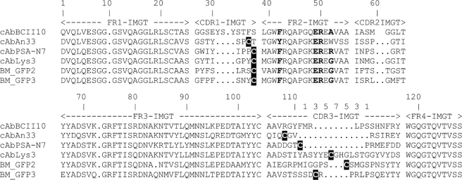
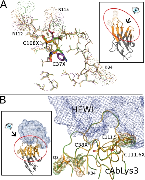
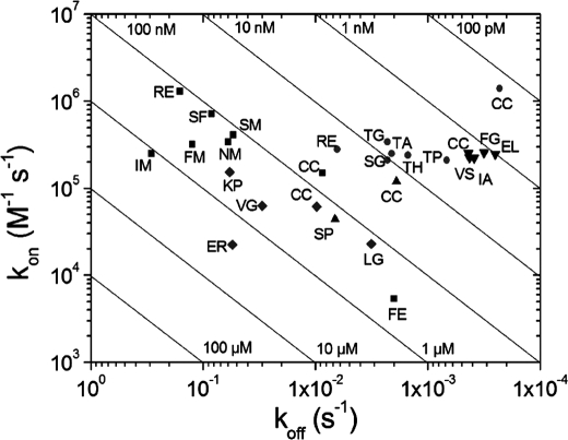
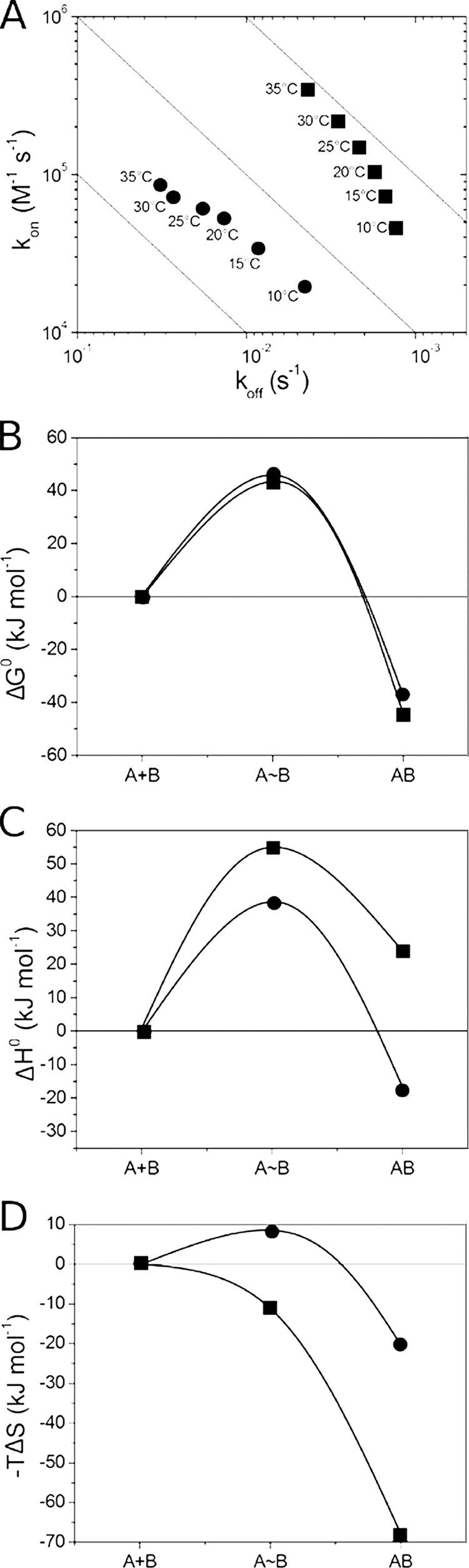
The antigen-binding fragment of functional heavy chain antibodies (HCAbs) in camelids comprises a single domain, named the variable domain of heavy chain of HCAbs (VHH). The VHH harbors remarkable amino acid substitutions in the framework region-2 to generate an antigen-binding domain that functions in the absence of a light chain partner. The substitutions provide a more hydrophilic, hence more soluble, character to the VHH but decrease the intrinsic stability of the domain. Here we investigate the functional role of an additional hallmark of dromedary VHHs, i.e. the extra disulfide bond between the first and third antigen-binding loops. After substituting the cysteines forming this interloop cystine by all 20 amino acids, we selected and characterized several VHHs that retain antigen binding capacity. Although VHH domains can function in the absence of an interloop disulfide bond, we demonstrate that its presence constitutes a net advantage. First, the disulfide bond stabilizes the domain and counteracts the destabilization by the framework region-2 hallmark amino acids. Second, the disulfide bond rigidifies the long third antigen-binding loop, leading to a stronger antigen interaction. This dual beneficial effect explains the in vivo antibody maturation process favoring VHH domains with an interloop disulfide bond.


Figures

FIGURE 1.
FIGURE 1.
Alignment of VHH amino acid sequences to universal VHH scaffold cAbBCII10 (31). The FRs, antigen-binding loops, CDRs, and amino acid numbering are according to IMGT. The hallmark amino acids in FR-2 that differentiate a VHH from VH are represented in bold and on a gray background. These VHH hallmark amino acids ((F/Y)ER(A/R/G)) are in VH highly conserved, hydrophobic (VGLW), and interact with the VL partner domain. The cysteines forming the VHH-characteristic interloop disulfide bond are highlighted in white lettering on a black background.
FIGURE 2.
FIGURE 2.
Structural model of cAbAbn33 and cAbLys3. A, thin line stick representation of the amino acid residues near the paratope of cAbAn33 (Protein Data Bank code 1YC7) (orange) superposed with two of the selected variants (cAbAn33 RE, green; cAbAn33 FE, purple). The interloop disulfide bond and the Cys-substituting residues are shown in thicker line stick representation colored by element, keeping the colors of the other amino acids of the VHH for the carbon atoms. The amino acids in the vicinity of cystine or its substitutions that differ in orientation are labeled and indicated by their surface contours. B, line presentation of cAbLys3 paratope (Protein Data Bank code 1MEL; orange) superposed on the cAbLys3 SP variant (green). The amino acids in the vicinity of cystine (Cys-38 and Cys-111.6) or its Ser-Pro substitutions that differ in orientation are labeled Q3, K84, and E111.5) and indicated by their surface contours. The interaction with the HEWL antigen (blue chicken wire representation) is also shown. Three-dimensional models of the WT and Cys mutant VHHs were generated using the ESyPred3D web server (38). Insets show the intact molecule and the eye view angle of the main picture.
FIGURE 3.
FIGURE 3.
RaPID plot for parental and variants of cAbAn33 (■), cAbPSA-N7 (●), cAbLys3 (▴), BM_GFP2 (▾), and BM_GFP3 (♦). The single letter code for the interloop disulfide bonded Cys or its amino acid substitutions are given for each VHH.
FIGURE 4.
FIGURE 4.
Thermodynamic characterization of interaction between cAbLys3 CC (■) or cAbLys3 SP (^) and HEWL. A, RaPID plot of the VHH-HEWL interaction at six different temperatures between 10 and 35 °C (temperature indicated adjacent to each data point in the plot). B–D, the reaction pathway of cAbLys3 CC or cAbLys3 SP interaction with HEWL. The changes in ΔG0, ΔH0, and −TΔS (B, C, and D, respectively) along the reaction coordinate are given for the initial unassociated state (A+B), the transition state (AB), and the antibody-antigen complex (AB).
FIGURE 5.
FIGURE 5.
Thermodynamic characterization of interaction between BM_GFP2 (CC (■), FG (●), IA (▴), EL (▾), or VS (♦)) and GFP along reaction pathway. The changes in ΔG0, ΔH0, and −TΔS (A, B, and C, respectively) along the reaction coordinate are given for the initial unassociated state (A+B), the transition state (AB), and the antibody-antigen complex (AB).
FIGURE 6.
FIGURE 6.
Fraction of unfolded cAbAn33 as function of temperature (A) or GdmCl concentration (B). Properly folded VHH at 35 °C or in a physiological buffer is unfolded by gradually increasing the temperature or the GdmCl concentration, respectively. The unfolding is followed spectrophotometrically, and the data are treated according to Saerens et al. (34). The open symbols in A and B (○ and □) are for the native cAbAn33 CC, and the filled symbols are for its variants (SM, ^; RE, ▴; FM, ▾; FE, ♦; SF, ◭; IM, ◮; NM, ■).

References

    1. Williams A. F., Barclay A. N. (1988) The immunoglobulin superfamily—domains for cell surface recognition. Annu. Rev. Immunol. 6, 381–405 - PubMed
    1. Lesk A. M., Chothia C. (1982) Evolution of proteins formed by β-sheets. II. The core of the immunoglobulin domains. J. Mol. Biol. 160, 325–342 - PubMed
    1. Lefranc M. P. (2004) IMGT-ONTOLOGY and IMGT databases, tools and web resources for immunogenetics and immunoinformatics. Mol. Immunol. 40, 647–660 - PubMed
    1. Abkevich V. I., Shakhnovich E. I. (2000) What can disulfide bonds tell us about protein energetics, function and folding: simulations and bioinformatics analysis. J. Mol. Biol. 300, 975–985 - PubMed
    1. Betz S. F. (1993) Disulfide bonds and the stability of globular proteins. Protein Sci. 2, 1551–1558 - PMC - PubMed

Publication types

LinkOut - more resources