Skp1 prolyl 4-hydroxylase of dictyostelium mediates glycosylation-independent and -dependent responses to O2 without affecting Skp1 stability
- PMID: 22128189
- PMCID: PMC3265880
- DOI: 10.1074/jbc.M111.314021
Skp1 prolyl 4-hydroxylase of dictyostelium mediates glycosylation-independent and -dependent responses to O2 without affecting Skp1 stability
Abstract
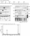
Cytoplasmic prolyl 4-hydroxylases (PHDs) have a primary role in O(2) sensing in animals via modification of the transcriptional factor subunit HIFα, resulting in its polyubiquitination by the E3(VHL)ubiquitin (Ub) ligase and degradation in the 26 S proteasome. Previously thought to be restricted to animals, a homolog (P4H1) of HIFα-type PHDs is expressed in the social amoeba Dictyostelium where it also exhibits characteristics of an O(2) sensor for development. Dictyostelium lacks HIFα, and P4H1 modifies a different protein, Skp1, an adaptor of the SCF class of E3-Ub ligases related to the E3(VHL)Ub ligase that targets animal HIFα. Normally, the HO-Skp1 product of the P4H1 reaction is capped by a GlcNAc sugar that can be subsequently extended to a pentasaccharide by novel glycosyltransferases. To analyze the role of glycosylation, the Skp1 GlcNAc-transferase locus gnt1 was modified with a missense mutation to block catalysis or a stop codon to truncate the protein. Despite the accumulation of the hydroxylated form of Skp1, Skp1 was not destabilized based on metabolic labeling. However, hydroxylation alone allowed for partial correction of the high O(2) requirement of P4H1-null cells, therefore revealing both glycosylation-independent and glycosylation-dependent roles for hydroxylation. Genetic complementation of the latter function required an enzymatically active form of Gnt1. Because the effect of the gnt1 deficiency depended on P4H1, and Skp1 was the only protein labeled when the GlcNAc-transferase was restored to mutant extracts, Skp1 apparently mediates the cellular functions of both P4H1 and Gnt1. Although Skp1 stability itself is not affected by hydroxylation, its modification may affect the stability of targets of Skp1-dependent Ub ligases.
Figures


Similar articles
-
Requirements for Skp1 processing by cytosolic prolyl 4(trans)-hydroxylase and α-N-acetylglucosaminyltransferase enzymes involved in O₂ signaling in dictyostelium.Biochemistry. 2011 Mar 15;50(10):1700-13. doi: 10.1021/bi101977w. Epub 2011 Feb 9. Biochemistry. 2011. PMID: 21247092 Free PMC article.
-
A cytoplasmic prolyl hydroxylation and glycosylation pathway modifies Skp1 and regulates O2-dependent development in Dictyostelium.Biochim Biophys Acta. 2010 Feb;1800(2):160-71. doi: 10.1016/j.bbagen.2009.11.006. Epub 2009 Nov 13. Biochim Biophys Acta. 2010. PMID: 19914348 Free PMC article. Review.
-
Prolyl hydroxylation- and glycosylation-dependent functions of Skp1 in O2-regulated development of Dictyostelium.Dev Biol. 2011 Jan 15;349(2):283-95. doi: 10.1016/j.ydbio.2010.10.013. Epub 2010 Oct 20. Dev Biol. 2011. PMID: 20969846 Free PMC article.
-
The Skp1 prolyl hydroxylase from Dictyostelium is related to the hypoxia-inducible factor-alpha class of animal prolyl 4-hydroxylases.J Biol Chem. 2005 Apr 15;280(15):14645-55. doi: 10.1074/jbc.M500600200. Epub 2005 Feb 10. J Biol Chem. 2005. PMID: 15705570
-
Evolutionary and functional implications of the complex glycosylation of Skp1, a cytoplasmic/nuclear glycoprotein associated with polyubiquitination.Cell Mol Life Sci. 2003 Feb;60(2):229-40. doi: 10.1007/s000180300018. Cell Mol Life Sci. 2003. PMID: 12678488 Free PMC article. Review.
Cited by
-
The aerotaxis of Dictyostelium discoideum is independent of mitochondria, nitric oxide and oxidative stress.Front Cell Dev Biol. 2023 Jun 15;11:1134011. doi: 10.3389/fcell.2023.1134011. eCollection 2023. Front Cell Dev Biol. 2023. PMID: 37397260 Free PMC article.
-
Chemical Synthesis of a Glycopeptide Derived from Skp1 for Probing Protein Specific Glycosylation.Chemistry. 2015 Aug 10;21(33):11779-87. doi: 10.1002/chem.201501598. Epub 2015 Jul 15. Chemistry. 2015. PMID: 26179871 Free PMC article.
-
Golgi UDP-GlcNAc:polypeptide O-α-N-Acetyl-d-glucosaminyltransferase 2 (TcOGNT2) regulates trypomastigote production and function in Trypanosoma cruzi.Eukaryot Cell. 2014 Oct;13(10):1312-27. doi: 10.1128/EC.00165-14. Epub 2014 Aug 1. Eukaryot Cell. 2014. PMID: 25084865 Free PMC article.
-
The E3 Ubiquitin Ligase Adaptor Protein Skp1 Is Glycosylated by an Evolutionarily Conserved Pathway That Regulates Protist Growth and Development.J Biol Chem. 2016 Feb 26;291(9):4268-80. doi: 10.1074/jbc.M115.703751. Epub 2015 Dec 30. J Biol Chem. 2016. PMID: 26719340 Free PMC article.
-
The Skp1 protein from Toxoplasma is modified by a cytoplasmic prolyl 4-hydroxylase associated with oxygen sensing in the social amoeba Dictyostelium.J Biol Chem. 2012 Jul 20;287(30):25098-110. doi: 10.1074/jbc.M112.355446. Epub 2012 May 30. J Biol Chem. 2012. PMID: 22648409 Free PMC article.
References
-
- Ward J. P. (2008) Oxygen sensors in context. Biochim. Biophys. Acta 1777, 1–14 - PubMed
-
- Kaelin W. G., Jr., Ratcliffe P. J. (2008) Oxygen sensing by metazoans: the central role of the HIF hydroxylase pathway. Mol. Cell 30, 393–402 - PubMed
-
- Schindler J., Sussman M. (1977) Ammonia determines the choice of morphogenetic pathways in Dictyostelium discoideum. J. Mol. Biol. 116, 161–169 - PubMed
Publication types
MeSH terms
Substances
Grants and funding
LinkOut - more resources
Full Text Sources
Molecular Biology Databases

