. 2012 Jan 6;287(2):1600-8.
doi: 10.1074/jbc.M111.312736. Epub 2011 Nov 29.

Brain-derived neurotrophic factor (BDNF) induces polarized signaling of small GTPase (Rac1) protein at the onset of Schwann cell myelination through partitioning-defective 3 (Par3) protein

Affiliations

Brain-derived neurotrophic factor (BDNF) induces polarized signaling of small GTPase (Rac1) protein at the onset of Schwann cell myelination through partitioning-defective 3 (Par3) protein

Chhavy Tep et al. J Biol Chem. .

Abstract

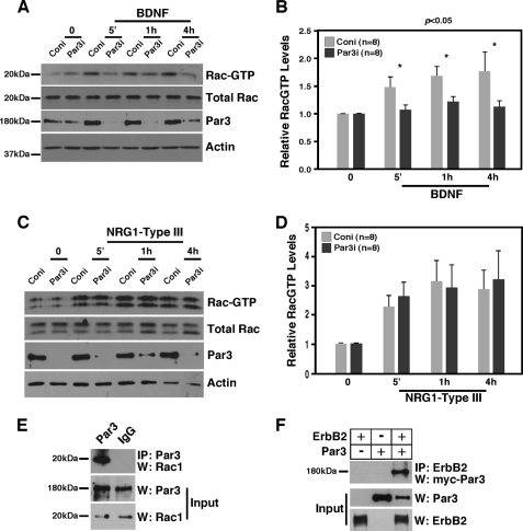
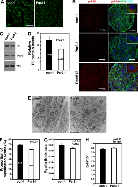
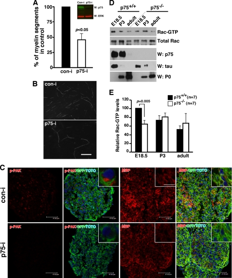
Brain-derived neurotrophic factor (BDNF) was shown to play a role in Schwann cell myelination by recruiting Par3 to the axon-glial interface, but the underlying mechanism has remained unclear. Here we report that Par3 regulates Rac1 activation by BDNF but not by NRG1-Type III in Schwann cells, although both ligands activate Rac1 in vivo. During development, active Rac1 signaling is localized to the axon-glial interface in Schwann cells by a Par3-dependent polarization mechanism. Knockdown of p75 and Par3 individually inhibits Rac1 activation, whereas constitutive activation of Rac1 disturbs the polarized activation of Rac1 in vivo. Polarized Rac1 activation is necessary for myelination as Par3 knockdown attenuates myelination in mouse sciatic nerves as well as in zebrafish. Specifically, Par3 knockdown in zebrafish disrupts proper alignment between the axon and Schwann cells without perturbing Schwann cell migration, suggesting that localized Rac1 activation at the axon-glial interface helps identify the initial wrapping sites. We therefore conclude that polarization of Rac1 activation is critical for myelination.


Figures

FIGURE 1.
FIGURE 1.
BDNF and NRG1-Type III activate Rac1 in sciatic nerves. A, RacGTP levels are their highest at E19.5 in rats and gradually decline during development. B, quantification of RacGTP levels in A. n = 6–8. C, asymmetric localization of phospho-PAK immunoreactivity in premyelinating Schwann cells in culture and in vivo. Note that phospho-PAK staining is along the side of Schwann cells that were in contact with DRG axons in culture. NF, neurofilament. Scale bar = 5 μm (left panels). Similarly, p-PAK immunoreactivity is found predominantly adjacent to axons in P3 sciatic nerves (right panel). Scale bar = 12 μm). The inset shows a high-magnification view. D, injection of TrkB-Fc and not control Fc into the sciatic nerves of P0 mice resulted in inhibition of Rac1 activation and P0 protein levels. Quantification is shown next to the figure. E, injection of ErbB3-Fc, and not control Fc, into the sciatic nerves of P0 mice resulted in inhibition of Rac1 activation and P0 protein levels. Quantification is shown next to the figure. F, BDNF activates Rac1 in primary Schwann cells. BDNF was added at 50 ng/ml for 1 h. Quantification is shown below the figures. G, NRG1-Type III activates Rac1 in primary Schwann cells. Membrane fractions (8 μg) from 293 cells expressing a vector or NRG1-Type III cDNA were added onto Schwann cells for 1 h. Quantification is shown below the figures. Relative RacGTP levels represent the RacGTP levels adjusted to total Rac1 level in each sample. For statistical analyses, Student's t test was used.
FIGURE 2.
FIGURE 2.
Par3 is necessary for BDNF-mediated but not for NRG1-Type III-mediated Rac1 activation in Schwann cell cultures. A, Par3 is necessary for BDNF-mediated Rac1 activation in Schwann cell cultures. Following infection with the retrovirus carrying Par3 RNAi (Par3i) or control RNAi (Coni), Schwann cells were treated with 50 ng/ml BDNF for the indicated period of time. Control Par3 Western blot analysis demonstrates the extent of Par3 knockdown. B, quantification of A. Relative RacGTP levels represent the RacGTP levels adjusted to total Rac1 level in each sample. For statistical analyses, Student's t test was used. C, knocking down Par3 in Schwann cells failed to inhibit Rac1 activation by NRG1-Type III. Following infection with the retrovirus carrying Par3 RNAi, Schwann cells were treated with 8 μg/ml NRG1-Type III for the indicated period of time. D, quantification of C. E, Par3 interacts with Rac1 in P3 sciatic nerves. Par3 was immunoprecipitated and blotted for Rac1. F, Par3 binds ErbB2 in 293T cells. Full-length Par3 and ErbB2 were transfected to 293T cells, and the resulting lysates were subjected to immunoprecipitation with ErbB2 and Western blot analysis with Par3. Control inputs are shown (5% of the lysates used for immunoprecipitation).
FIGURE 3.
FIGURE 3.
Selective knockdown of Par3 among Schwann cells in vivo results in disruption of polarized Rac1 activation and attenuation in myelination. A, representative images of the retrovirus-infected sciatic nerves. GFP signals indicate the circular myelin sheath formed by the Schwann cells that were infected with the retroviruses. The retroviruses carry GFP, so infected myelinating Schwann cells appear green in doughnut shapes. Scale bar = 15 μm. B, knockdown of Par3 in vivo inhibits polarized Rac1 activation in Schwann cells. Note that p-PAK immunoreactivity is polarized within Schwann cells (inset). Although p-PAK immunoreactivity is detected throughout the cross-section of the sciatic nerve from the control virus-infected mice, it is significantly reduced in the nerves that were infected with shRNA-carrying retroviruses of Par3. In contrast, RacV12 infection increased p-PAK immunoreactivity throughout the sciatic nerve and disrupted its polarization. Scale bars = 45–47 μm. The inset shows a high-magnification view. C, knockdown of Par3 in vivo resulted in reduction in P0 protein levels. Par3i, Par3 RNAi; Coni, control RNAi. Also shown is the control Western blot analysis, Par3, and tau. D, quantification of P0 proteins from C. For statistical analyses, Student's t test was used. E, representative EM images of the retrovirus-infected sciatic nerves at P4. Scale bar = 10 μm. F, quantification of the proportion of the axons that were myelinated. Note that there were four different mice that had been injected with the virus. For statistical analyses, Student's t test was used. G, myelin thickness was reduced with Par3 knockdown. H, the g ratio was increased with Par3 knockdown.
FIGURE 4.
FIGURE 4.
Knockdown of pard3 in zebrafish larvae resulted in abnormal Schwann cell wrapping and a significant reduction in mbp expression. A, pard3 MO1 reduces Pard3 protein levels and inhibits RacGTP levels in zebrafish larvae. Embryos at 4 dpf were deyolked, and proteins were extracted for RacGTP assays and control Western blot analyses. B–G, lateral views at the level of the trunk of 4 dpf larvae hybridized for mbp. In control larvae (B and C), mbp is expressed in stripes associated with ventral motor roots (B, asterisks) and along the posterior lateral line nerve (C, arrow). In pard3 MO1- (D and E) and MO2 (F and G)-injected larvae, mbp expression is lost at the ventral roots (asterisks) and the posterior lateral line nerve (arrows), whereas it is still evident in the spinal cord (sc, brackets). H and I, lateral views at the level of the trunk of 4 dpf Tg(olig2:EGFP);Tg(sox10:mRFP) larvae. Motor axons are green, and Schwann cells are red. In a control larva (H), Schwann cells wrap motor axons (arrows), whereas in a MO1 injected larva (I), Schwann cells failed to align along the axon, forming a loose association with the axon. The MO-injected larva also has a deficit of axon wrapping by oligodendrocytes, which are marked by RFP expression, in the spinal cord.
FIGURE 5.
FIGURE 5.
P75 signals from Schwann cells activate Rac1 and promote myelination. A, knockdown of p75 in rat Schwann cells inhibited the extent of myelination in culture. Also shown are the control Westerns for the p75 knockdown with RNAi. For statistical analyses, Student's t test was used. B, representative images of MBP staining of the cocultures. Scale bar = 10 μm. C, knockdown of p75 among Schwann cells reduced the extent of p-PAK and MBP immunoreactivity. Note that we are using the same image from Fig. 3B for the control shRNA-infected nerves. Scale bars = 45–47 μm. D, P75 is necessary for Rac1 activation in vivo. RacGTP levels are reduced in p75−/− mice compared with those in p75+/+ at E18.5. As controls, the protein levels of p75, tau, and P0 are also shown. Note that there is a reduction in tau protein levels, reflecting a reduction in DRG fibers. E, quantification of RacGTP from D.

Similar articles

Cited by

References

    1. Goldstein B., Macara I. G. (2007) The PAR proteins. Fundamental players in animal cell polarization. Dev. Cell 13, 609–622 - PMC - PubMed
    1. Chan J. R., Jolicoeur C., Yamauchi J., Elliott J., Fawcett J. P., Ng B. K., Cayouette M. (2006) The polarity protein Par-3 directly interacts with p75NTR to regulate myelination. Science 314, 832–836 - PubMed
    1. Ng B. K., Chen L., Mandemakers W., Cosgaya J. M., Chan J. R. (2007) Anterograde transport and secretion of brain-derived neurotrophic factor along sensory axons promote Schwann cell myelination. J. Neurosci. 27, 7597–7603 - PMC - PubMed
    1. Chen X., Macara I. G. (2005) Par-3 controls tight junction assembly through the Rac exchange factor Tiam1. Nat. Cell Biol. 7, 262–269 - PubMed
    1. Nishimura T., Kato K., Yamaguchi T., Fukata Y., Ohno S., Kaibuchi K. (2004) Role of the PAR-3-KIF3 complex in the establishment of neuronal polarity. Nat. Cell Biol. 6, 328–334 - PubMed

Publication types

MeSH terms