

Inhibition of experimental myopia by a dopamine agonist: different effectiveness between form deprivation and hyperopic defocus in guinea pigs
- PMID: 22128230
- PMCID: PMC3224832
Inhibition of experimental myopia by a dopamine agonist: different effectiveness between form deprivation and hyperopic defocus in guinea pigs
Abstract
Purpose: The dopamine (DA) system in the retina is critical to normal visual development as lack of retinal DA signaling may contribute to myopic development. The involvement of DA in myopic development is complex and may be different between form deprivation and hyperopic defocus. This study evaluated effects of a non-selective DA receptor agonist, apomorphine (APO) on refractive development in guinea pigs treated with form deprivation or hyperopic defocus.
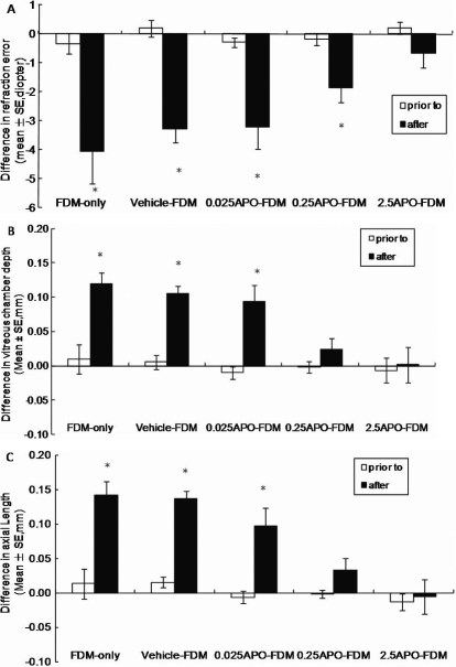
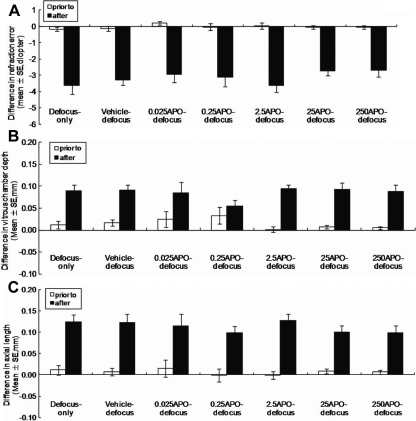
Methods: APO was subconjunctivally injected daily for 11 days in form-deprived (0.025 to 2.5 ng/µl) and defocused (0.025 to 250 ng/µl) eyes. Changes in ocular biometry and retinal concentration of DA and its metabolites (DOPAC) were measured in the 2 animal models to assess the level of DA involvement in each of the models (the less the change, the lower the involvement).
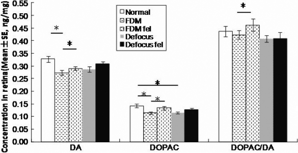
Results: Similar myopic degree was induced in both the deprived and defocused eyes (-4.06 D versus -3.64 D) at 11 days of the experiment. DA and DOPAC levels were reduced in the deprived eyes but did not change significantly in the defocused eyes compared to the fellow and normal control eyes. A subconjunctival injection of APO daily for 11 days at concentrations ranged from 0.025 to 2.5 ng/µl inhibited form deprivation myopia in a concentration-dependent manner. By contrast, the APO treatment ranged from 0.025 to 250 ng/µl did not effectively inhibit the defocus-induced myopia and the associated axial elongation.
Conclusions: DA signaling may play a more critical role in form deprivation myopia than in defocus-induced myopia, raising a question whether the mechanisms of DA signaling are different under these two types of experimental myopia.
Figures
Similar articles
-
The Role of Dopamine in Emmetropization Modulated by Wavelength and Temporal Frequency in Guinea Pigs.Invest Ophthalmol Vis Sci. 2021 Sep 2;62(12):20. doi: 10.1167/iovs.62.12.20. Invest Ophthalmol Vis Sci. 2021. PMID: 34546324 Free PMC article.
-
Dopamine Receptor Subtypes Mediate Opposing Effects on Form Deprivation Myopia in Pigmented Guinea Pigs.Invest Ophthalmol Vis Sci. 2018 Sep 4;59(11):4441-4448. doi: 10.1167/iovs.17-21574. Invest Ophthalmol Vis Sci. 2018. PMID: 30193315
-
Myopia induced by flickering light in guinea pig eyes is associated with increased rather than decreased dopamine release.Mol Vis. 2017 Sep 29;23:666-679. eCollection 2017. Mol Vis. 2017. PMID: 28966549 Free PMC article.
-
Temporal integration of visual signals in lens compensation (a review).Exp Eye Res. 2013 Sep;114:69-76. doi: 10.1016/j.exer.2013.02.014. Epub 2013 Mar 5. Exp Eye Res. 2013. PMID: 23470505 Free PMC article. Review.
-
An updated view on the role of dopamine in myopia.Exp Eye Res. 2013 Sep;114:106-19. doi: 10.1016/j.exer.2013.02.007. Epub 2013 Feb 19. Exp Eye Res. 2013. PMID: 23434455 Review.
Cited by
-
Alteration of EIF2 Signaling, Glycolysis, and Dopamine Secretion in Form-Deprived Myopia in Response to 1% Atropine Treatment: Evidence From Interactive iTRAQ-MS and SWATH-MS Proteomics Using a Guinea Pig Model.Front Pharmacol. 2022 Jan 28;13:814814. doi: 10.3389/fphar.2022.814814. eCollection 2022. Front Pharmacol. 2022. PMID: 35153787 Free PMC article.
-
Update in myopia and treatment strategy of atropine use in myopia control.Eye (Lond). 2019 Jan;33(1):3-13. doi: 10.1038/s41433-018-0139-7. Epub 2018 Jun 11. Eye (Lond). 2019. PMID: 29891900 Free PMC article. Review.
-
The Role of Retinal Dysfunction in Myopia Development.Cell Mol Neurobiol. 2023 Jul;43(5):1905-1930. doi: 10.1007/s10571-022-01309-1. Epub 2022 Nov 24. Cell Mol Neurobiol. 2023. PMID: 36427109 Free PMC article. Review.
-
Changes in Intrinsically Photosensitive Retinal Ganglion Cells, Dopaminergic Amacrine Cells, and Their Connectivity in the Retinas of Lid Suture Myopia.Invest Ophthalmol Vis Sci. 2024 Sep 3;65(11):8. doi: 10.1167/iovs.65.11.8. Invest Ophthalmol Vis Sci. 2024. PMID: 39230992 Free PMC article.
-
SWATH Based Quantitative Proteomics Reveals Significant Lipid Metabolism in Early Myopic Guinea Pig Retina.Int J Mol Sci. 2021 Apr 29;22(9):4721. doi: 10.3390/ijms22094721. Int J Mol Sci. 2021. PMID: 33946922 Free PMC article.
References
-
- Rohrer B, Iuvone PM, Stell WK. Stimulation of dopaminergic amacrine cells by stroboscopic illumination or fibroblast growth factor (bFGF, FGF-2) injections: possible roles in prevention of form-deprivation myopia in the chick. Brain Res. 1995;686:169–81. - PubMed
-
- Wallman J, Winawer J. Homeostasis of eye growth and the question of myopia. Neuron. 2004;43:447–68. - PubMed
-
- Rymer J, Wildsoet CF. The role of the retinal pigment epithelium in eye growth regulation and myopia: a review. Vis Neurosci. 2005;22:251–61. - PubMed
-
- Rada JA, Wiechmann AF. Melatonin receptors in chick ocular tissues: implications for a role of melatonin in ocular growth regulation. Invest Ophthalmol Vis Sci. 2006;47:25–33. - PubMed
-
- Mao J, Liu S, Qin W, Li F, Wu X, Tan Q. Levodopa inhibits the development of form-deprivation myopia in guinea pigs. Optom Vis Sci. 2010;87:53–60. - PubMed
Publication types
MeSH terms
Substances
LinkOut - more resources
Full Text Sources
Other Literature Sources
Research Materials
Miscellaneous