. 2011:17:2847-55.
Epub 2011 Nov 9.

Changes of the ocular surface and aquaporins in the lacrimal glands of rabbits during pregnancy

Affiliations

Changes of the ocular surface and aquaporins in the lacrimal glands of rabbits during pregnancy

Chuanqing Ding et al. Mol Vis. 2011.

Abstract

Purpose: To test the hypotheses that pregnancy represents a physiologic condition that is associated with dry eye symptoms, and the expression of aquaporin 4 (AQP4) and AQP5 are altered in the lacrimal gland (LG) from term pregnant rabbits.

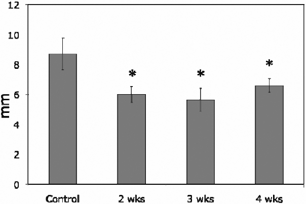
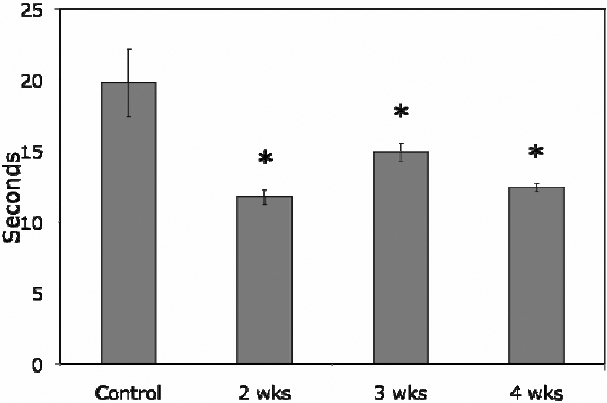
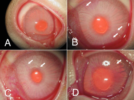
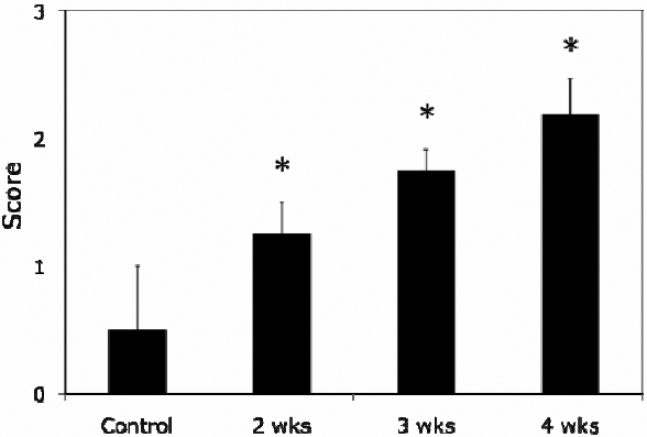
Methods: Schirmer's test, tear break-up time (BUT), and Rose Bengal staining were used to evaluate ocular surface health. LG were obtained from term pregnant rabbits and age-matched female control rabbits and then processed for laser capture microdissection (LCM), real time RT-PCR, western blot, and immunofluorescence for the detection and quantification of mRNA and proteins of AQP4 and AQP5.

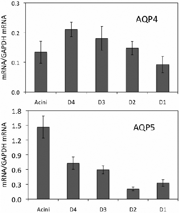
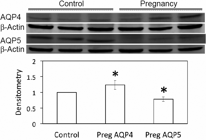
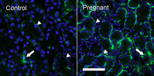
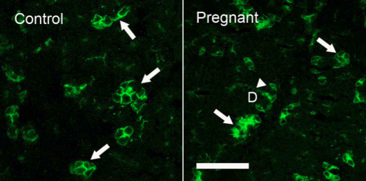
Results: Pregnant rabbits demonstrated typical clinical symptoms of dry eye, including decreased Schirmer score and BUT as well as increased Rose Bengal staining of cornea. In term pregnant rabbits, mRNA for AQP5 from whole LG was significantly lower than that of control rabbits, while mRNA for AQP4 was not. Levels of mRNA for AQP4 and AQP5 underwent significant changes in acini and epithelial cells from specific duct segments during pregnancy. Western blot from whole LG lysates demonstrated that expression of AQP4 was 24% more abundant in term pregnant rabbits while AQP5 was 22% less when compared to control rabbits respectively. At term pregnancy, AQP4 immunoreactivity (AQP4-IR) was increased in acini while its intensity remained the same in ducts. AQP5-IR was present in both apical and basolateral membranes of acinar cells in normal control and pregnant rabbits, while ductal cells in pregnant rabbits also showed significant amount of AQP5-IR.

Conclusions: The data presented here demonstrated significant dry eye symptoms in pregnant rabbits. Our data also showed altered expressions of AQP4 and AQP5 during pregnancy and suggested that these changes may contribute to the altered LG secretion and dry eye symptoms during pregnancy.


Figures

Figure 1
Figure 1
Schirmer tests of normal control and pregnant rabbits. Schirmer score was 8.69±1.07 mm in normal control rabbits while the scores were reduced to 6±0.54 mm at 2 weeks’ pregnancy, 5.63±0.77 mm at 3 weeks, and 6.59±0.45 mm at 4 weeks pregnancy, with the score at each time point of pregnancy was significantly lower than that of normal controls (p<0.05, as indicated by *). Data are presented as mean±standard error of the mean (SEM).
Figure 2
Figure 2
BUT was 19.79±2.39 s in normal control rabbits and was reduced to 11.75±0.51 s at 2 weeks’ pregnancy, 14.9±0.65 s at 3 weeks, and 12.43±0.31 s at 4 weeks pregnancy. Each time point of pregnancy was significantly lower than that of normal control (p<0.05, as indicated by *). Data are presented as mean±SEM.
Figure 3
Figure 3
Representative images of Rose Bengal staining of rabbit corneas. The test was unremarkable in normal control rabbits (A), but starting at 2 weeks of pregnancy, punctate staining (arrows) was observed in the corneas of many rabbits (B). At 3 weeks of pregnancy, the rabbit cornea typically shows a moderate staining (C, arrows). Moderate to severe staining was found in approximately 50% of pregnant rabbits at 4 weeks pregnancy (D). As shown in panel D, one rabbit’s cornea showed prominent punctate staining in every quadrant of the cornea.
Figure 4
Figure 4
Grading of Rose Bengal staining. The scores increased from 0.5±0.5 in controls to 1.25±0.25 of 2 weeks’ pregnancy, and 1.75±0.16 of 3 weeks, and 2.19±0.28 of 4 weeks. Data are presented as mean±SEM.
Figure 5
Figure 5
Real-time RT–PCR of AQP4 and AQP5 from LCM samples of term pregnant rabbits. Table 2 compares these results to that of normal rabbit controls. mRNA levels for AQP4 was the lowest in interlobar duct, rather than acini as observed in control rabbits [5], and its level was significantly lower in all duct segments except interlobular duct (p<0.05). AQP5 mRNA was the highest in acini and was significantly lower (p<0.05) than that of normal control rabbits [5]. However, mRNA abundance was significantly increased in intralobular, interlobular, and interlobar ducts (p<0.05), while no change was found in intralobar duct. D4: intralobular duct. D3: interlobular duct. D2: intralobar duct. D1: interlobar duct. Data are presented as mean±SEM of 3 animals.
Figure 6
Figure 6
Western blots of AQP4 and AQP5 from whole LG homogenates. AQP4 was significantly increased in LG from pregnant rabbits, while AQP5 was decreased (p<0.05). β-Actin was used as loading control. Data are representative images of at least 3 different animals each.
Figure 7
Figure 7
Immunofluorescence of AQP4-IR. Control: AQP4-IR was observed on the basolateral sides of acinar and duct cells, with duct (arrow) showing much stronger AQP4-IR than acini (arrowheads), results in accordance with our previous reports [5,6]. Pregnant: Acini in pregnant rabbits showed substantially stronger basolateral staining (arrowheads) than control, whereas AQP4-IR in ducts (arrow) appeared to be similar to that of control. In both images, DAPI was used to stain nuclei as bright blue to demonstrate the morphologic profiles of acini and ducts. Scale bar=50 μm.
Figure 8
Figure 8
Immunofluorescence of AQP5-IR. Control: AQP5-IR was present in both basolateral and apical membranes of acinar cells, and distributed among acini in a “mosaic” pattern, with some acini and/or acinar cells demonstrating much stronger AQP5-IR (arrows) than the rest of acini/acinar cells. However, little AQP5-IR was detected in duct cells. These results were similar to our previous reports [5,6]. Pregnant: as in control LG, AQP5-IR was also present in a “mosaic” pattern with a similar intensity and distribution pattern (arrows). However, in contrast to control animals, ductal cells also exhibited a significant amount of AQP5-IR (arrowhead). D=duct. Scale bar=50 μm.

Similar articles

Cited by

References

    1. Pflugfelder SC, Tseng S, Sanabria O, Kell H, Garcia C, Felix C, Feuer W, Reis B. Evaluation of subjective assessments and objective diagnostic tests for diagnosing tear-film disorders known to cause ocular irritation. Cornea. 1998;17:38–56. - PubMed
    1. Wong J, Ding C, Yiu S, Smith R, Goodwin T, Schechter JE. An Epidemiological Study of Pregnancy and Dry Eye. Ocul Surf. 2004;3:S127.
    1. Sullivan DA. Tearful relationships? Sex, hormones, the lacrimal gland, and aqueous-deficient dry eye. Ocul Surf. 2004;2:92–123. - PubMed
    1. Ding C, Chang N, Fong YC, Wang Y, Trousdale MD, Mircheff AK, Schechter JE. Interacting Influences of Pregnancy and Corneal Injury on Rabbit Lacrimal Gland Immunoarchitecture and Function. Invest Ophthalmol Vis Sci. 2006;47:1368–75. - PubMed
    1. Ding C, Parsa L, Nandoskar P, Zhao P, Wu K, Wang Y. Duct system of the rabbit lacrimal gland: Structural characteristics and its role in lacrimal secretions. Invest Ophthalmol Vis Sci. 2010;51:2960–7. - PMC - PubMed

Publication types

MeSH terms

LinkOut - more resources