. 2011 Nov;3(1 Suppl):S7-S19.
doi: 10.1177/1758834011422556.

An overview of the c-MET signaling pathway

Affiliations

An overview of the c-MET signaling pathway

Shawna Leslie Organ et al. Ther Adv Med Oncol. 2011 Nov.

Abstract

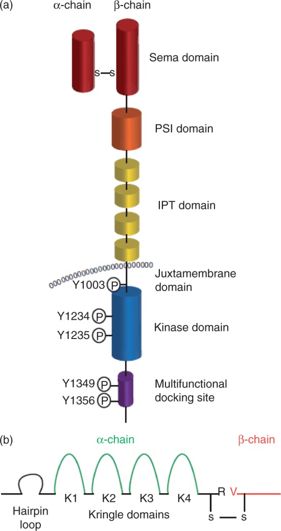
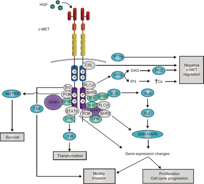
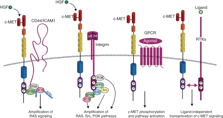
c-MET is a receptor tyrosine kinase that, after binding with its ligand, hepatocyte growth factor, activates a wide range of different cellular signaling pathways, including those involved in proliferation, motility, migration and invasion. Although c-MET is important in the control of tissue homeostasis under normal physiological conditions, it has also been found to be aberrantly activated in human cancers via mutation, amplification or protein overexpression. This paper provides an overview of the c-MET signaling pathway, including its role in the development of cancers, and provides a rationale for targeting the pathway as a possible treatment option.

Keywords: MET; c-MET; cancer; hepatocyte growth factor (HGF); receptor tyrosine kinase; signaling.


Figures

Figure 1.
Figure 1.
Domain structure of c-MET and hepatocyte growth factor (HGF). (a) The c-MET receptor is formed by proteolytic processing of a common precursor into a single-pass, disulphide-linked α/β heterodimer. The extracellular portion of c-MET is composed of three domain types. The N-terminal 500 residues fold to form a large semaphorin (Sema) domain, which encompasses the whole α-subunit and part of the β-subunit. The plexin–semaphorin–integrin (PSI) domain follows the Sema domain, spans approximately 50 residues and includes four disulphide bonds. This domain is connected to the transmembrane helix via four immunoglobulin–plexin–transcription (IPT) domains, which are related to immunoglobulin-like domains. Intracellularly, the c-MET receptor contains a tyrosine kinase catalytic domain flanked by distinctive juxtamembrane and carboxy-terminal sequences. This portion of c-MET contains the catalytic tyrosines Y1234 and Y1235, which positively modulate enzyme activity, while the juxtamembrane tyrosine 1003 negatively regulates c-MET by recruiting the ubiquitin ligase casitas B-lineage lymphoma (c-CBL). The multifunctional docking site in the C-terminal tail contains tyrosines Y1349 and Y1356, which recruit several transducers and adaptors when c-MET is active. (b) The c-MET ligand, hepatocyte growth factor (HGF), is secreted by mesenchymal cells as a single-chain, biologically inert precursor and is converted into its bioactive form when extracellular proteases cleave the bond between Arg494 and Val495. The mature form of HGF consists of an α- and β-chain, which are held together by a disulphide bond. The α-chain contains an N-terminal hairpin loop followed by four kringle domains (80 amino acid double-looped structures formed by three internal disulphide bridges), K1–4. The β-chain is homologous to the serine proteases of the blood-clotting cascade, but lacks any proteolytic activity. Adapted from Comoglio et al. [2008].
Figure 2.
Figure 2.
c-MET signaling adaptors and mediators. When the tyrosines within the multifunctional docking site become phosphorylated they recruit signaling effectors, including the adaptor proteins growth factor receptor-bound protein 2 (GRB2), src homology 2 domain-containing (SHC), v-crk sarcoma virus CT10 oncogene homolog (CRK) and CRK-like (CRKL); the effector molecules phosphatidylinositol 3-kinase (PI3K), phospholipase Cγ (PLCγ) and SRC, the src homology 2 domain-containing 5' inositol phosphatase SHIP-2, and the signal transducer and activator of transcription STAT3. In addition, unique to c-MET is its association with the adaptor protein GRB2-associated binding protein 1 (GAB1), a multi-adaptor protein that, once bound to and phosphorylated by c-MET, creates binding sites for more downstream adaptors. GAB1 can bind either directly to c-MET or indirectly, through GRB2. The downstream response to c-MET activation relies on stereotypical signaling modulators common to many receptor tyrosine kinases. For activation of the mitogen-activated protein kinase (MAPK) cascades, c-MET activation stimulates the activity of the rat sarcoma viral oncogene homolog (RAS) guanine nucleotide exchanger son of sevenless (SOS) via binding with SHC and GRB2 leading to the activation of RAS. This leads to the indirect activation of v-raf murine sarcoma viral oncogene homolog B1 (RAF) kinases, which can subsequently activate MAPK effector kinase (MEK), and finally MAPK, which can then translocate to the nucleus to activate the transcription factors responsible for regulating a large number of genes, including those involved in cell proliferation, cell motility and cell cycle progression. SHP2 can also link c-MET signaling to the MAPK cascade, as sequestration of SHP2 to GAB1 is responsible for extending the duration of MAPK phosphorylation. The p85 subunit of PI3K can bind either directly to c-MET or indirectly through GAB1, which then signals through AKT/protein kinase B. This axis is primarily responsible for the cell survival response to c-MET signaling. Transformation downstream of the c-MET receptor is mediated by the phosphorylation of Janus kinase 1 (JNK), which occurs via binding to CRK. STAT3 has also been implicated in transformation. The direct binding of STAT3 to c-MET results in STAT3 phosphorylation, dimerization and its translocation to the nucleus. This has been shown to result in tubulogenesis and invasion. However, other reports have found that, although STAT3 is required for c-MET-mediated tumorigenesis, it has no effect on proliferation, invasion or branching morphogenesis. Cellular migration is also mediated downstream of c-MET by focal adhesion kinase (FAK), which is localized to cellular adhesion complexes. FAK is activated through phosphorylation by SRC family kinases, which have been shown to directly associate with c-MET. The c-MET–SRC–FAK interaction leads to cell migration and the promotion of anchorage-independent growth. Negative regulation of the c-MET receptor is crucial for its tightly controlled activity. The Y1003 site, located in the juxtamembrane domain, is a negative regulatory site for c-MET signaling that acts by recruiting c-CBL. Regulation of c-MET signaling is also accomplished via its binding to various protein tyrosine phosphatases (PTPs). These PTPs modulate c-MET signaling by dephosphorylation of either the tyrosines in the c-MET kinase or the docking site. Finally, binding of PLCγ to c-MET results in the activation of protein kinase C (PKC), which can then negatively regulate c-MET receptor phosphorylation and activity. Independently of PKC activation, an increase in intracellular calcium levels can also lead to negative c-MET regulation. Adapted from Trusolino et al. [2010] and Birchmeier et al. [2003]. DAG, diacylglycerol; HGF, hepatocyte growth factor; IP3, inositol triphosphate; PIP3, phosphatidylinositol (3,4,5)-triphosphate.
Figure 3.
Figure 3.
c-MET transactivation. The potency and endurance of c-MET-triggered pathways is secured by a network of upstream signaling co-receptors that physically associate with c-MET at the cell surface. c-MET membrane partners can then amplify and/or diversify c-MET-dependent biochemical inputs and translate them into meaningful (and specific) biological outcomes. The v6 splice variant of the hyaluronan receptor CD44 links c-MET signaling to the actin cytoskeleton via the growth factor receptor-bound protein 2 (GRB2) and the ezrin, radixin, moesin (ERM) family of proteins in order to recruit son of sevenless (SOS), which then amplifies RAS-ERK signaling. Intercellular adhesion molecule 1 (ICAM-1) can substitute for CD44v6 as a co-receptor for c-MET in CD44v6 knockout mice, resulting in similar c-MET pathway activation. c-MET binding to integrin α6β4 creates a supplementary docking platform for the binding of signaling adaptors, leading to specific enhancement of phosphatidylinositol 3-kinase (PI3K), RAS and SRC activation. c-MET can also be activated by G-protein coupled receptors (GPCRs), although the functional outcome of this interaction is not well characterized. Crosstalk between c-MET and other receptor tyrosine kinases (RTKs) has also been studied in great depth because of its potential importance in the development of resistance to cancer therapeutics. Examples of these RTKs include the semaphorin receptors, the epidermal growth factor receptor (EGFR) family of receptors, the recepteur d'origine nantais (RON), platelet-derived growth factor receptor (PDGFR) and Axl; the list continues to grow. Adapted from Trusolino et al. [2010] and Corso et al. [2005] HGF, hepatocyte growth factor; SHC, src homolgy 2 domain-containing; SHP2, src homology 2 domain-containing phosphatase 2.

References

    1. Abounader R., Reznik T., Colantuoni C., Martinez-Murillo F., Rosen E.M., Laterra J. (2004) Regulation of c-Met-dependent gene expression by PTEN. Oncogene 23: 9173–9182 - PubMed
    1. Bachleitner-Hofmann T., Sun M.Y., Chen C.T., Tang L., Song L., Zeng Z., et al. (2008) HER kinase activation confers resistance to MET tyrosine kinase inhibition in MET oncogene-addicted gastric cancer cells. Mol Cancer Ther 7: 3499–3508 - PubMed
    1. Basilico C., Arnesano A., Galluzzo M., Comoglio P.M., Michieli P. (2008) A high affinity hepatocyte growth factor-binding site in the immunoglobulin-like region of Met. J Biol Chem 283: 21267–21277 - PMC - PubMed
    1. Bean J., Brennan C., Shih J.Y., Riely G., Viale A., Wang L., et al. (2007) MET amplification occurs with or without T790M mutations in EGFR mutant lung tumors with acquired resistance to gefitinib or erlotinib. Proc Natl Acad Sci USA 104: 20932–20937 - PMC - PubMed
    1. Benvenuti S., Lazzari L., Arnesano A., Li Chiavi G., Gentile A., Comoglio P.M. (2011) Ron kinase transphosphorylation sustains MET oncogene addiction. Cancer Res 71: 1945–1955 - PubMed

LinkOut - more resources