. 2012 Jan;18(1):111-23.
doi: 10.1261/rna.029454.111. Epub 2011 Nov 29.

The long noncoding RNA Vax2os1 controls the cell cycle progression of photoreceptor progenitors in the mouse retina

Affiliations

The long noncoding RNA Vax2os1 controls the cell cycle progression of photoreceptor progenitors in the mouse retina

Nicola Meola et al. RNA. 2012 Jan.

Abstract

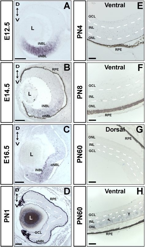
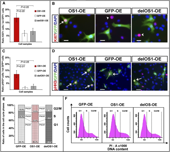
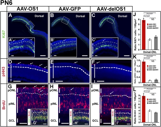
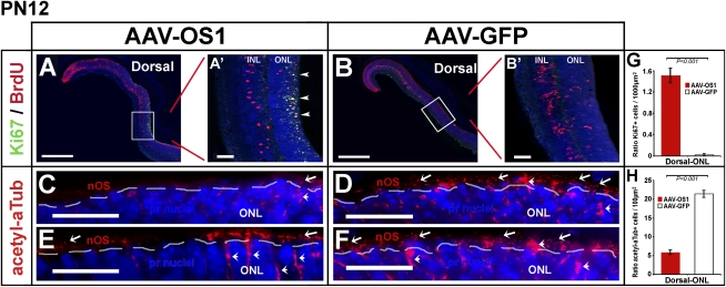
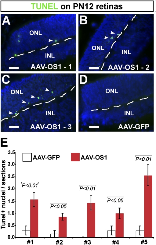
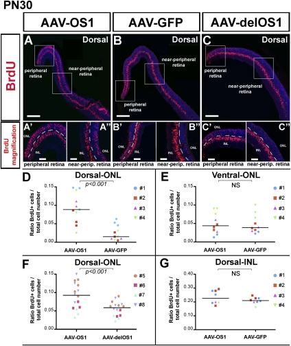
Long noncoding RNAs (lncRNAs) are emerging as regulators of many basic cellular pathways. Several lncRNAs are selectively expressed in the developing retina, although little is known about their functional role in this tissue. Vax2os1 is a retina-specific lncRNA whose expression is restricted to the mouse ventral retina. Here we demonstrate that spatiotemporal misexpression of Vax2os1 determines cell cycle alterations in photoreceptor progenitor cells. In particular, the overexpression of Vax2os1 in the developing early postnatal mouse retina causes an impaired cell cycle progression of photoreceptor progenitors toward their final committed fate and a consequent delay of their differentiation processes. At later developmental stages, this perturbation is accompanied by an increase of apoptotic events in the photoreceptor cell layer, in comparison with control retinas, without affecting the proper cell layering in the adult retina. Similar results are observed in mouse photoreceptor-derived 661W cells in which Vax2os1 overexpression results in an impairment of the cell cycle progression rate and cell differentiation. Based on these results, we conclude that Vax2os1 is involved in the control of cell cycle progression of photoreceptor progenitor cells in the ventral retina. Therefore, we propose Vax2os1 as the first example of lncRNA that acts as a cell cycle regulator in the mammalian retina during development.


Figures

FIGURE 1.
FIGURE 1.
Vax2os1 expression pattern in the embryonic and postnatal mouse retina by RNA in situ hybridization (ISH). (A–C) Vax2os1 is expressed in the ventral areas of the mouse embryonic retina from stages E12.5–E16.5. (D–F) In the postnatal stages, Vax2os1 is not detected by RNA ISH, while in the adult retina, Vax2os1 is expressed only in the outer nuclear layer of the ventral retina (arrowheads) (G,H). All pictures derive from C57/Bl6 mouse retinas except for A and C, in which albino CD1 mice were used. (oNBL) Outer neuroblastic retina; (iNBL) inner neuroblastic retina; (GCL) ganglion cell layer; (RPE) retinal pigment epithelium; (L) lens; (ONL) outer nuclear layer; (INL) inner nuclear layer; (D) dorsal; (V) ventral. Note that the borders of the retinal cell layers of postnatal and adult retinas are marked by dotted lines. Magnifying bars, 100 μm (A–D); 40 μm (E–H).
FIGURE 2.
FIGURE 2.
Vax2os1 overexpression impairs the cell cycle of 661W cells. Characterization of the 661W cells undergoing a differentiation protocol for 48 h (see text for details). (A,B) Ki67 fluorescence immunostaining. (C,D) Phospho-histone-H3, pHH3, fluorescence immunostaining. In A and C, the histograms show that the Vax2os1-transfected cells (OS1-OE) are characterized by an increase in the number of cells positive to Ki67 (A) or pHH3 (C) compared with the pool of control transfected cells (GFP-OE and delOS1-OE; see text for details). In B, the arrow indicates one Vax2os1-transfected cell expressing Ki67; arrowheads indicate cells expressing only Ki67 in control experiments. In D, arrows indicate cells expressing both pHH3 and GFP in either the OS1-OE or the GFP-OE and delOS1-OE cells. (E,F) Cell cycle analysis with propidium iodide (PI). (E) Histograms show the distribution (in percentage) of the single cycle phases for the OS1-OE cells (middle bar) in comparison with the GFP-OE (left bar) and delOS1-OE control (right bar) cell pools. Differences between the GFP-OE and delOS1-OE were not significant. (F) Flow cytometric DNA content analysis of the OS1-OE cells and GFP-OE and delOS1-OE control cells stained with PI. (A,C) Statistical P-values from likelihood ratio test for the negative binomial; (E) statistical P-value from χ2 test. Magnifying bars, 20 μm (B); 40 μm (D). (A,C) Data are represented as means ± SEM, n = 4.
FIGURE 3.
FIGURE 3.
Vax2os1 overexpression in the retinal postnatal stages affects the correct proliferation of retinal progenitor cells. AAVs overexpressing Vax2os1 and the two control constructs were injected into the dorsal retinal areas of PN1 mice. (A–C) Ki67 fluorescent immunostaining (green signal) observed in the PN6 dorsal retina areas. (A′–C′) Higher magnifications of the areas boxed in panels A–C. (Arrows) Ki67-positive cells in the prospective outer nuclear layer (pONL) of the AAV-OS1-injected retinas and the control construct-injected retinal areas. (D–F) pHH3 fluorescent immunostaining (in red) on the PN6 dorsal retina areas. (Arrows) pHH3-positive cells in the pONL of AAV-OS1-injected retinas and the control-injected retinal areas. (G–I) BrdU fluorescent detection in the PN6 dorsal retina areas. The BrdU was administered 24 h before killing the animals. There is an increase of BrdU-positive cells in the pONL of the AAV-OS1-injected retinas compared with the control-injected retinal areas (arrows). (G′–I′) Lower magnifications of panels G–H showing the results of coimmunostaining for BrdU and Ki67. The retinal cells positive to both the markers (in yellow) are present in the pINL and pONL in both the AAV-OS1- and control-injected retinas. However, there is an evident increase in the number of the BrdU/Ki67-positive cells in the pONL of the AAV-OS1-injected retinas compared with the control-injected retinal areas (arrowheads). Note that the lower limit of the pONL is indicated with a dotted line. (J–L) Counts of the Ki67 (J), pHH3 (K), and BrdU (L) positive cells in the dorsal ONL of the injected retinas at PN6. Areas of 30 μm × 20 μm were used for manual counts. For each animal, multiple retinal areas were analyzed. (pINL) Prospective inner nuclear layer; (GCL) ganglion cell layer. Magnifying bars, 200 μm (A–C); 40 μm (A′–C′,G–I′); 100 μm (D–F). (J–L) Values are means ± SEM (n = 4) and P-values derived from likelihood ratio test for negative binomial.
FIGURE 4.
FIGURE 4.
Alteration in the differentiation of photoreceptor cells following Vax2os1 overexpression. (A,B) Fluorescence coimmunostaining for BrdU and Ki67 in the dorsal areas of PN1-injected retinas at PN12. (A′,B′) Higher magnifications of the boxed areas in panels A and B, respectively. There is an increase in the number of the cells positive to both BrdU and Ki67 (arrowheads in A′) in the outer nuclear layer (ONL) of the AAV-OS1-injected retinas in comparison with the AAV-GFP-injected retinal areas. (C–F) Fluorescence immunostaining for acetylated α-tubulin (acetyl-αTub) in the dorsal retinal areas of the injected mice at PN12. There is a decrease in the number of the acetyl-αTub-positive photoreceptor cells (arrows) in the AAV-OS1-injected, as compared with the control-injected retinas. The dotted lines delimit the border between the nOS and the ONL. Note that acetyl-αTub also stains the cytoplasm of Müller glial cells (arrowheads). (G,H) Counts of the Ki67-positive cells (G) and acetyl-aTub-positive photoreceptor outer segments (H) in the dorsal ONL of the injected retinas at PN12. Areas of 160 μm × 40 μm (G) and 80 μm × 5 μm (H) were used for manual counts. For each animal, multiple retinal areas were analyzed. Magnifying bars, 40 μm (A′,B′); 20 μm (C–F). (G,H) Values are means ± SEM (n = 3) and P-values derived from likelihood ratio test for negative binomial.
FIGURE 5.
FIGURE 5.
Increase of apoptosis at PN12 in the photoreceptor layer after Vax2os1-overexpression. (A–D) Detection of programmed cell death by terminal deoxynucleotidyl transferase dUTP nick end labeling method (TUNEL, in green, arrowheads) in the dorsal retinas of the AAV-OS1- (A–C) and AAV-GFP-injected (D) mice. There is an increase in the number of the TUNEL-positive cells in the AAV-OS1- versus control-injected retinas (D). (E) Quantitative assessment/section of the number of TUNEL-positive nuclei in the dorsal retinal regions analyzed. A significant increase of the TUNEL-positive cells was observed in the AAV-OS1-injected retinas over control-injected retinas in each of the five animals analyzed. Magnifying bars, 20 μm. (E) Results are means ± SEM from seven technical replicates and P-values derived from a Student's t-test.
FIGURE 6.
FIGURE 6.
Vax2os1 action is restricted to the photoreceptor cells. (A–C) The BrdU assay on PN30 dorsal retinal areas of mice injected at PN1. Two pulses of BrdU were performed, at PN5 and PN6, before sacrifice. (A′–C′,A″–C″) Higher magnifications of, respectively, the peripheral and near-peripheral retinal areas boxed in panels A–C. (D–G) Counts of BrdU-positive cells in the near-peripheral retinal areas of injected animals. In each panel, the identity of the animal analyzed is indicated. Areas of 80 μm × 40 μm were used for manual BrdU counts. For each animal, multiple retinal areas were analyzed. (y-axis) The total cell number value indicates the number of photoreceptor nuclei within the aforementioned areas. Magnifying bars, 200 μm (A–C); 40 μm (A′–C″). The horizontal line in each graphic represents the Grand Median as derived from the GraphPad Prism program. (D–G) Data are means ± SEM, n = 4, and P-values derived from likelihood ratio test for negative binomial.

References

    1. Alexiades MR, Cepko C 1996. Quantitative analysis of proliferation and cell cycle length during development of the rat retina. Dev Dyn 205: 293–307 - PubMed
    1. Alfano G, Vitiello C, Caccioppoli C, Caramico T, Carola A, Szego MJ, McInnes RR, Auricchio A, Banfi S 2005. Natural antisense transcripts associated with genes involved in eye development. Hum Mol Genet 14: 913–923 - PubMed
    1. Alfano G, Conte I, Caramico T, Avellino R, Arnò B, Pizzo MT, Tanimoto N, Beck SC, Huber G, Dollé P, et al. 2011. Vax2 regulates retinoic acid distribution and cone opsin expression in the vertebrate eye. Development 138: 261–271 - PubMed
    1. Allocca M, Mussolino C, Garcia-Hoyos M, Sanges D, Iodice C, Petrillo M, Vandenberghe LH, Wilson JM, Marigo V, Surace EM, et al. 2007. Novel adeno-associated virus serotypes efficiently transduce murine photoreceptors. J Virol 81: 11372–11380 - PMC - PubMed
    1. Al-Ubaidi MR, Font RL, Quiambao AB, Keener MJ, Liou GI, Overbeek PA, Baehr W 1992. Bilateral retinal and brain tumors in transgenic mice expressing simian virus 40 large T antigen under control of the human interphotoreceptor retinoid-binding protein promoter. J Cell Biol 119: 1681–1687 - PMC - PubMed

Publication types