. 2012 Jan;18(1):65-76.
doi: 10.1261/rna.02809212. Epub 2011 Nov 29.

RNA single strands bind to a conserved surface of the major cold shock protein in crystals and solution

Affiliations

RNA single strands bind to a conserved surface of the major cold shock protein in crystals and solution

Rolf Sachs et al. RNA. 2012 Jan.

Abstract

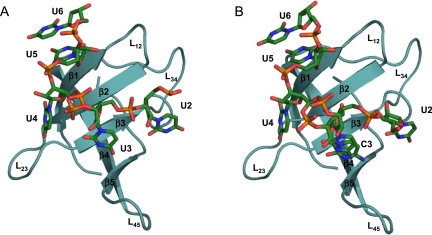
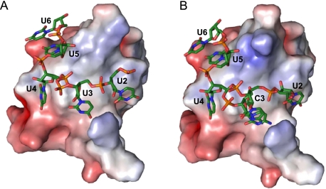
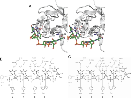
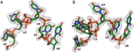
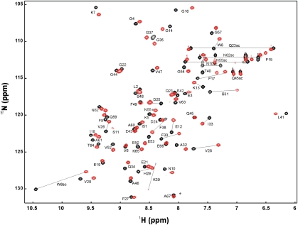
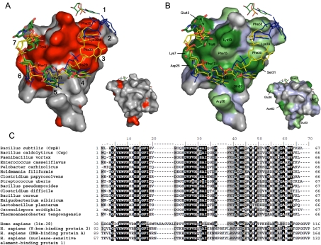
Bacterial cold shock proteins (CSPs) regulate the cellular response to temperature downshift. Their general principle of function involves RNA chaperoning and transcriptional antitermination. Here we present two crystal structures of cold shock protein B from Bacillus subtilis (Bs-CspB) in complex with either a hexanucleotide (5'-UUUUUU-3') or heptanucleotide (5'-GUCUUUA-3') single-stranded RNA (ssRNA). Hydrogen bonds and stacking interactions between RNA bases and aromatic sidechains characterize individual binding subsites. Additional binding subsites which are not occupied by the ligand in the crystal structure were revealed by NMR spectroscopy in solution on Bs-CspB·RNA complexes. Binding studies demonstrate that Bs-CspB associates with ssDNA as well as ssRNA with moderate sequence specificity. Varying affinities of oligonucleotides are reflected mainly in changes of the dissociation rates. The generally lower binding affinity of ssRNA compared to its ssDNA analog is attributed solely to the substitution of thymine by uracil bases in RNA.


Figures

FIGURE 1.
FIGURE 1.
Crystal structures of Bs-CspB in complex with RNA. The ribbon (Bs-CspB) and stick (RNA) representation of the complexes (A) Bs-CspB·rU6 and (B) Bs-CspB·rC7 are labeled according to the secondary structure elements, the loops (L), and the nucleotides.
FIGURE 2.
FIGURE 2.
Electrostatic surface potential of the Bs-CspB ligand binding site. The electrostatic potential is projected onto the molecular surface of the protein of (A) Bs-CspB·rU6 and (B) Bs-CspB·rC7. The electrostatic potential was calculated with APBS (Baker et al. 2001) for pH 7.5, with a range from −5 kT (red) to 5 kT (blue).
FIGURE 3.
FIGURE 3.
Stacking and polar interactions between Bs-CspB and the ligands (A,C) rC7 and (B) rU6. (A) A stereo representation of the Bs-CspB molecule with the longest observed nucleotide sequence of rC7 is shown. The backbone of the protein molecule is depicted as gray object; protein groups involved in stacking interactions and water-mediated or direct hydrogen bonding are colored according to the CPK scheme with the exception of carbon, which is in gray. The RNA is colored according to the CPK scheme with the exception of carbon, which is green. Direct hydrogen bonds between protein and RNA are shown as dotted lines. In a schematic depiction of ligand binding the RNA strands (B) rU6 and (C) rC7 are depicted in black and groups of Bs-CspB in dark gray (bb, protein backbone). The longest observed nucleotide sequence of rC7 is presented. A group belonging to a symmetry-related Bs-CspB is displayed in light gray. Stacking interactions between aromatic sidechains and RNA bases are shown as solid gray lines. Hydrogen bonds are displayed as dotted lines. The numbers of the contact subsites for individual bases are given at the bottom, using the numbering scheme introduced earlier (Max et al. 2006).
FIGURE 4.
FIGURE 4.
RNA ligands (A) rU6 and (B) rC7 surrounded by their electron density. The ssRNA is colored according to the CPK scheme with the exception of carbon, which is green. Their composite omit electron densities are contoured corresponding to 1.0 σ. (A) Five of six uridine nucleotides of rU6 could be built into the electron density. The phosphodiester group connecting nucleotides 2 and 3 adopts a double conformation. (B) Nucleotides U2 to U6 of rC7 could be built into the density. Nucleotide C3 and the phosphodiester group of the following nucleotide exist in two conformations. The occupancy of U2 is 0.5 such that a sterical clash with the C3 nucleotide which would collide in one of its two conformations is avoided.
FIGURE 5.
FIGURE 5.
2D 1H/15N HSQC spectrum of free (black) and rC7-bound (red) Bs-CspB in 20 mM HEPES, 50 mM NaCl, pH 7.5, 10% D2O at 20°C. The spectrum of rC7-bound Bs-CspB was recorded at a 1.5-fold molar excess of rC7.
FIGURE 6.
FIGURE 6.
Ligand binding surface of Bs-CspB and its phylogenetic conservation. (A) The interaction of the ssRNA rC7 with Bs-CspB as monitored by NMR titration experiments is depicted and compared with published CSP ssDNA complexes. For the surface representation, the crystal structure of the Bs-CspB·rC7 complex has been used. Residues standing out from chemical shift changes of the backbone amides (ΔδMW > 0.13 ppm) are shown in red. Amino acid residues stacked against RNA or DNA bases in the crystals of Bs-CspB·rC7, Bs-CspB·rU6, and the ssDNA complexes Bs-CspB·dT6 and Bc-Csp·dT6 as well as the backbone of Lys39 are labeled. The numbers of the respective contact subsites for individual bases are also indicated. The ssRNA rC7 of the present work is presented as sticks and colored according to the CPK scheme with the exception of carbon, which is in green. The symmetry-related C3 base contacting subsite 1 is also indicated. The ssDNA molecule of the Bs-CspB·dT6 complex is depicted in blue and the ssDNA molecule of the Bc-CspB·dT6 complex in yellow. The small surface depicts the protein backside. (B) Same representation as in A with the Bs-CspB surface colored according to sequence conservation. Most residues forming the ligand interaction site are conserved at the level of at least 75% identity (dark green) or similarity (light green). Invariant regions which originate from the protein backbone are colored light blue. (C) Sequence alignment of bacterial CSP (upper) and CSD from human proteins (lower panel). Residues that are conserved at a level of at least 75% identity or similarity are highlighted in black or gray, respectively.

Similar articles

Cited by

References

    1. Bae W, Jones PG, Inouye M 1997. CspA, the major cold shock protein of Escherichia coli, negatively regulates its own gene expression. J Bacteriol 179: 7081–7088 - PMC - PubMed
    1. Bae W, Xia B, Inouye M, Severinov K 2000. Escherichia coli CspA-family RNA chaperones are transcription antiterminators. Proc Natl Acad Sci 97: 7784–7789 - PMC - PubMed
    1. Baker NA, Sept D, Joseph S, Holst MJ, McCammon JA 2001. Electrostatics of nanosystems: Application to microtubules and the ribosome. Proc Natl Acad Sci 98: 10037–10041 - PMC - PubMed
    1. Brandi A, Pon CL, Gualerzi CO 1994. Interaction of the main cold shock protein CS7.4 (CspA) of Escherichia coli with the promoter region of hns. Biochimie 76: 1090–1098 - PubMed
    1. Brandi A, Spurio R, Gualerzi CO, Pon CL 1999. Massive presence of the Escherichia coli ‘major cold-shock protein’ CspA under non-stress conditions. EMBO J 18: 1653–1659 - PMC - PubMed

Publication types

LinkOut - more resources