. 2012 Sep;16(9):2049-59.
doi: 10.1111/j.1582-4934.2011.01500.x.

Activation of the prolyl-hydroxylase oxygen-sensing signal cascade leads to AMPK activation in cardiomyocytes

Affiliations

Activation of the prolyl-hydroxylase oxygen-sensing signal cascade leads to AMPK activation in cardiomyocytes

Hong Yan et al. J Cell Mol Med. 2012 Sep.

Abstract

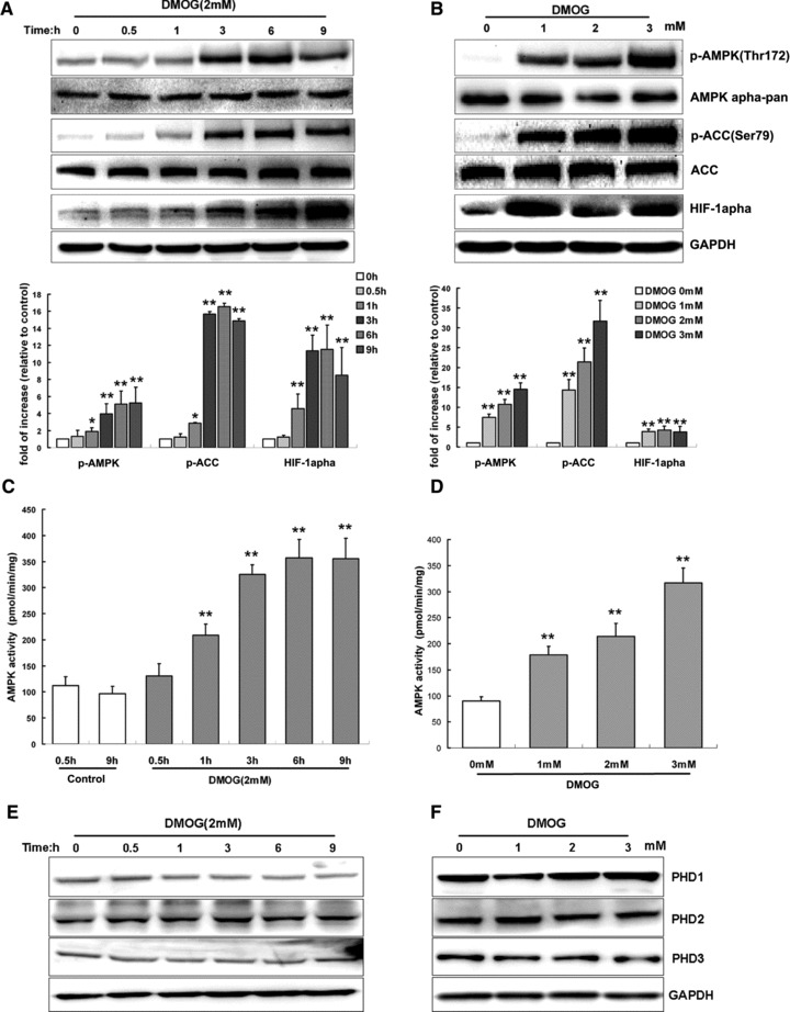
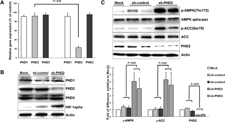
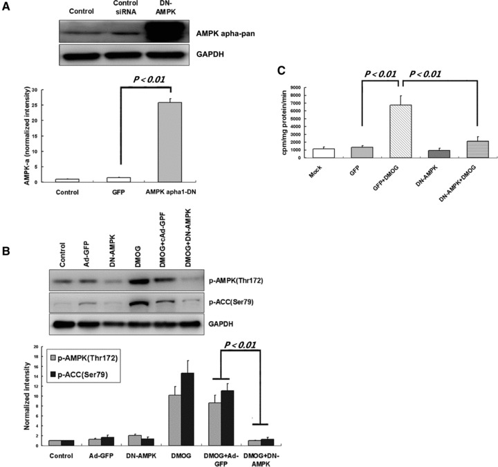
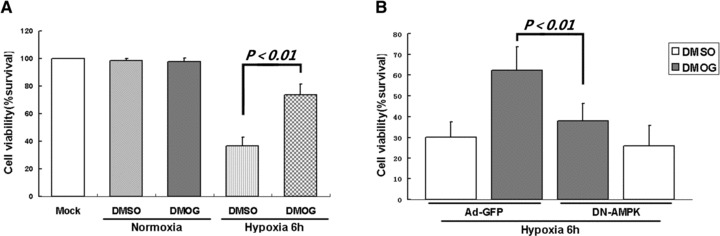
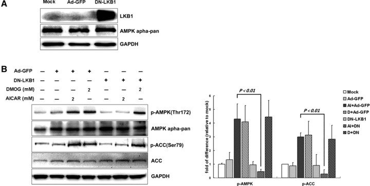
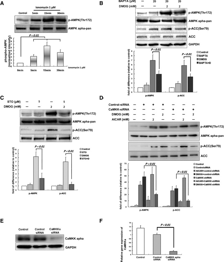
The proline hydroxylase domain-containing enzymes (PHD) act as cellular oxygen sensors and initiate a hypoxic signal cascade to induce a range of cellular responses to hypoxia especially in the aspect of energy and metabolic homeostasis regulation. AMP-activated protein kinase (AMPK) is recognized as a major energetic sensor and regulator of cardiac metabolism. However, the effect of PHD signal on AMPK has never been studied before. A PHD inhibitor (PHI), dimethyloxalylglycine and PHD2-specific RNA interference (RNAi) have been used to activate PHD signalling in neonatal rat cardiomyocytes. Both PHI and PHD2-RNAi activated AMPK pathway in cardiomyocytes effectively. In addition, the increased glucose uptake during normoxia and enhanced myocyte viability during hypoxia induced by PHI pretreatment were abrogated substantially upon AMPK inhibition with an adenoviral vector expressing a dominant negative mutant of AMPK-α1. Furthermore, chelation of intracellular Ca2+ by BAPTA, inhibition of calmodulin-dependent kinase kinase (CaMKK) with STO-609, or RNAi-mediated down-regulation of CaMKK α inhibited PHI-induced AMPK activation significantly. In contrast, down-regulation of LKB1 with adenoviruses expressing the dominant negative form did not affect PHI-induced AMPK activation. We establish for the first time that activation of PHD signal cascade can activate AMPK pathway mainly through a Ca(2+)/CaMKK-dependent mechanism in cardiomyocytes. Furthermore, activation of AMPK plays an essential role in hypoxic protective responses induced by PHI.


Figures

Fig 1
Fig 1
PHIs activate AMPK activity in cardiomyocytes in a time- and dose-dependent manner. Cardiomyocytes were treated with DMOG (2 mM) for indicated time periods (A, C, E) or different dose of DMOG for 3 hrs (B, D, F). For A and B, representative immunoblots together with densitometric analysis of four experiments are shown. Target protein phosphorylation has been normalized to the expression of the total protein. For C and D, AMPK activity was measured and shown as pmol/min./mg of protein. For E and F, representative immunoblots of PHD1, 2, 3 under same DMOG-treated conditions were shown. Each bar represents the mean ± S.D. for three independent assays in duplicate. **P < 0.01 versus control; *P < 0.05 versus control.
Fig 2
Fig 2
PHD2 knockdown leads to AMPK activation in cardiomyocyte. Cardiomyocytes were infected with adenoviruses for scrambled shRNA or PHD2 shRNA for 26 hrs, and then were collected for molecular analysis. (A) Real-time PCR for PHD1, 2, 3 (n = 3 independent experiments) shows fold change of mRNA. Values represent the mean ± S.D. (B and C) Representative immunoblots together with densitometric analysis (mean ± S.D.) of four independent experiments. AMPK and ACC phosphorylation has been normalized to the expression of total protein, respectively.
Fig 3
Fig 3
DMOG stimulates glucose uptake via AMPK activation. (A) Cardiomyocytes were exposed to 30 MOI of Ad-GFP or 20 MOI of DN-AMPK adenovirus for 24 hrs, hence, an equal transfection efficacy above 95% was obtained; cells were then harvested for Western blot analysis. (B) After adenovirus infection, cardiomyocytes were then exposed to 2 mM DMOG for three hrs before immunoblot assay. Representative immunoblots together with densitometric analysis (mean ± S.D.) of four independent experiments are shown. AMPK and ACC phosphorylation has been normalized to GAPDH expression. (C) After infection, cells were further incubated with DMOG (2 mM) or vehicle (DMSO) for 8 hrs. Then, 2-[3H]deoxy-D-glucose was added to culture media for 15 min., and glucose uptake was measured 3H accumulation rate which normalized to protein content. Each column represents means ± S.D. for three independent assays in duplicate.
Fig 4
Fig 4
AMPK activation is critical for PHI-induced hypoxic protection in cardiomyocytes. (A) Cardiomyocytes were incubated with DMOG (2 mM) or vehicle (DMSO) for 12 hrs, then subjected to hypoxic condition for six hrs. (B) Cardiomyocytes were infected with Ad-GFP or DN-AMPK constructs for 24 hrs, then exposed to DMOG (2 mM) or vehicle (DMSO) for 12 hrs, and subjected to hypoxia for 6 hrs. Cell viability was determined by CCK-8 assay. Data represent mean ± S.D. of three independent experiments in duplicate, and the cell survival ratio was expressed as a percentage of the control.
Fig 5
Fig 5
LKB1 is not required for PHI-induced AMPK activation in cardiomyocytes. (A) Cardiomyocytes were exposed for 24 hrs to 30 MOI of Ad-GFP or 40 MOI of DN-LKB1 adenovirus, after that, cells had been harvested for Western blot analysis. (B) After adenovirus infection with Ad-GFP or DN-LKB1 for 24 hrs, cardiomyocytes were then further exposed to 2 mM DMOG or vehicle (DMSO) for 3 hrs before immunoblot assay. Representative immunoblots together with densitometric analysis (mean ± S.D.) of four independent experiments are shown. AMPK and ACC phosphorylation has been normalized to the total protein expression, respectively.
Fig 6
Fig 6
CaMKKα is the major AMPK kinase upon PHI stimulation in cardiomyocytes. (A) Cardiomyocytes were stimulated with ionomycin (1 μM) for the indicated times. (B) Cardiomyocytes were pretreated with BAPTA-AM (20 μM, 30 min.) and subsequently stimulated with DMOG (2 mM) for three hrs. (C) Cardiomyocytes were preincubated with STO-609 (5 μM) for 30 min. before DMOG stimulation for three hrs; cells were lysed and subjected to immunoblotting. (D) Cardiomyocytes were incubated with transfection reagent only [polybrene (10 μg/ml), control] or with lentiviral shRNA vector targeted to rat CaMKKα or lentiviral control-shRNA (1 μg/30-mm dish) for four days. After that, cells were incubated with DMOG (2 mM), AICAR (2 mM) or DMSO for three hrs, and harvested for Western blot analysis. Representative blots and densitometry data (means ± S.D.) of four independent experiments for each treatment are shown. Cardiomyocytes were infected with lentiviral CaMKKα shRNA vector (1 μg/30-mm dish, 96 hrs) or control-shRNA or incubated with transfection reagent only (polybrene (10 μg/ml), control]. Representative immunoblot of CaMKKα (E) and graph of real-time PCR for CaMKKα (F) are shown. Values represent the mean ± S.D., n = 3 independent experiments. AMPK and ACC phosphorylation has been normalized to the total protein expression, respectively.

Similar articles

Cited by

References

    1. Bruick RK, McKnight SL. A conserved family of prolyl-4-hydroxylases that modify HIF. Science. 2001;294:1337–40. - PubMed
    1. Li SH, Shin DH, Chun YS, et al. A novel mode of action of YC-1 in HIF inhibition: stimulation of FIH-dependent p300 dissociation from HIF-1 alpha. Mol Cancer Ther. 2008;7:3729–38. - PubMed
    1. Ivan M, Kondo K, Yang HF, et al. HIF alpha targeted for VHL-mediated destruction by proline hydroxylation: implications for O-2 sensing. Science. 2001;292:464–8. - PubMed
    1. Kuznetsova AV, Meller J, Schnell PO, et al. von Hippel-Lindau protein binds hyperphosphorylated large subunit of RNA polymerase II through a proline hydroxylation motif and targets it for ubiquitination. Proc Natl Acad Sci U S A. 2003;100:2706–11. - PMC - PubMed
    1. Cummins EP, Berra E, Comerford KM, et al. Prolyl hydroxylase-1 negatively regulates I kappa B kinase-beta, giving insight into hypoxia-induced NF kappa B activity. Proc Natl Acad Sci U S A. 2006;103:18154–9. - PMC - PubMed

Publication types

MeSH terms

LinkOut - more resources